International Journal of Scientific & Engineering Research, Volume 4, Issue 6, June-2013 112
ISSN 2229-5518
Study of Optimal Classifiers based on
Computational Intelligence techniques for the
Diagnosis of Lung Cancer
Abstract— In this paper a new classification algorithm is proposed for the diagnosis of Lung cancer. In order to develop algorithm 39 CT scan images of patients have been considered consisting of Benign Tumor , Malignant Tumor and Normal Lung CT Scan iimage.W ith a view to extract features from the CT scan images after image processing , an algorithm is developed which proposes two-dimensional discrete cosine Transform domain coefficients in addition to Average, Standard Deviation, Entropy, Contrast, Correlation, Energy, Homogeneity. The suitability of classifiers based on Multilayer Perceptron (MLP) Neural Network is explored with the optimization of their respective parameters in view of reduction in time as well as space complexity. A separate Cross-Validation dataset is used for proper evaluation of the proposed classification algorithm with respect to important performance measures, such as MSE and classification accuracy. The Average Classification Accuracy of MLP Neural Network comprising of one hidden layers with 7 PE’s organized in a typical topology is found to be superior (100 %) for Training . Finally, optimal algorithm has been developed on the basis of the best classifier performance. The algorithm will provide an effective alternative to traditional method of Lung CT image analysis for deciding the tumor in lung is Benign or Malignant.
Index Terms—Optimal classifer,MLP, Computational Intellignce for diagnosis of lung cancer, Diagnosis of lung cancer by computational intelligence technique,Optimal classifiers for lung cancer,Neural Network ,Lung CT images,Cross validation for lung cancer.
—————————— ——————————
Cancer is a petrifying disease, death-dealing disease. The sufferer alone can know the torment it causes.
There are many types of cancers. Lung cancer is one of the most common and deadly diseases in the world. The Inci- dence, Lung cancer is on Second Top and the Highest in death rate . It is a dreaded cancer disease for the human death.
These patients are not confirmed with cancer & treated wrongly in early stages due to lack of experts, clinical inter- preters . The delay in detection, false diagnosis by experts, lack of experts in small towns, costly diagnosis are some of the reasons to these hapless victims for increase in death rate.
To mitigate their sufferings, an expert Lung cancer diag- nosis Computation Intelligence system has been developed where experts could get second opinion for the confirmation of the disease in its early, curable stage.
In this paper optimal classifier based on Computational Intelligence techniques for the diagnosis of Lung Cancer has been developed.
After regrious training & retraining of the classifier, it is cross validated & tested on the basis of many performance matrix.
Use of the optimal classifier based on Computational In- telligence techniques results in more accurate and reliable diagnosis of lung cancer disease. To elevate the plight of poor patients, our optimal classifier will prove to be a major boon.
Our system will help in diagnosis of lung cancer disease in its early stage, consequently the survival rate of patient can be pro-longed with affordable low cost treatment, medication etc etc.
The proposed algorithm provides classification using clas- sifiers based on Multi-layer Perceptron neural network ap- proach and tested on the Lung CT scan images comprising of features extracted using 2D DCT domain co-efficient .
Collected Lung CT images are in .jpg format. By using image processing & cropping the region of interest (ROI) the 128 fea- tures are extracted .
IJSER © 2013 http://www.ijser.org
International Journal of Scientific & Engineering Research, Volume 4, Issue 6, June-2013 113
ISSN 2229-5518
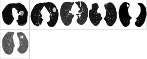
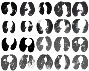
Fig. 1 Few Samples of input processed images of lung.
(Above lung images are of Benign,Malignant and Normal types)
Each Lung CT image is represented by a feature vector, F; which is comprised of 128 different parameters. The dataset contains 39 instances (exemplars) for three different classifica- tion.. The classifier based on neural network is trained from the training dataset, where a feature vector is mapped on to a particular class or name of the Lung disease. The neural net-
that processes the input image resulting in 2D discrete WHT domain coefficients in addition to Average, Standard Devia- tion, Entropy, Contrast, Correlation, Energy, Homogeneity and Shape descriptor. Here class and shape descriptor are symbolic or qualitative, whereas all other parameters are nu- meric-valued or quantitative. The values obtained were ex- ported to spreadsheet.
Neural Networks: Neuro Solutions (NeuroDimensions, Inc. USA) 5.0 was used to implement various NN based clas- sifiers on lung image which is represented by a Feature Vec- tor containing 128 different elements.
MLP based classifier were explored and studied with re- spect to the performance measures.
MSE (Mean Square Error):
The formula for the mean squared error is:
P N
work learns from data (training exemplars) and the connec- tion weights and biases are estimated as a result of this learn-
∑∑ (d
![]()
MSE = j = 0 i = 0
ij − yij )
(5)
ing. After training of the neural network, its connection
weights are frozen and latter; it is tested on a different da-
NP
Where P = number of output processing elements, N =
taset, which was never presented to the neural network. Here,
number of exemplars in the data set,
yij = neural network
this dataset is known as a cross-validation (CV) dataset. The performance of the classifier based on neural network is eval- uated on the basis of some metrics, such as, MSE, NMSE,
output for exemplar i at processing element j, output for exemplar i at processing element j.
NMSE (Normalized Mean Square Error):
dij = desired
Classification Accuracy and Confusion Matrix. In this work,
the prototype model of the classifier is developed with a view
to discriminate between 3 different lung diseases. However,
The normalized mean squared error is defined by the fol-
lowing formula:
P N MSE
the proposed algorithm can be easily applied for classification of more than 3 lung diseases provided that one has enough![]()
NMSE =
N N
N d 2 − d
computational resources. The feature vector, which is to be
extracted from the separated ROI of Lung image, is as fol-
lows.
F = [DCT1, DCT 2, DCT 3, ..., DCT 128, Average, Standard
P
∑
j = 0
∑ ij
i = 0
∑

i = 0
N
ij
(6)
Deviation, Entropy, Contrast, Correlation, Energy, Homoge- neity, Shape];
Where DCT 1, DCT 2, DCT3, ..., DCT 128 denote the two- dimensional discrete Cosine transform domain coefficients.
When working with large images, normal image pro- cessing techniques may sometimes break down, because the images can either be too large to load into memory, or else they can be loaded into memory but then be too large to pro- cess. To avoid these problems, block-processing approach is used, where one can process large images incrementally: reading, processing, and finally writing the results back to disk, one region at a time. In block-processing, an image, a block size, and a function handle are specified and then the input image is divided into blocks of the specified size. Later, all blocks are processed using the function handle one block at a time, and then the results are assembled into an output image. For lung images, block size of 16x16 is used for opti- mal results as compared to block size of 4x4 and 8x8.
An environment, accessible from MATLAB R2010b
(Mathworks Inc., USA) is used to implement the algorithm
Where P = number of output processing elements, N =
number of exemplars in the data set, MSE = mean square er- ror, dij = desired output for exemplar i at processing element j.
A confusion matrix is a simple methodology for display- ing the classification results of a network. The confusion ma- trix is defined by labeling the desired classification on the rows and the predicted classifications on the columns. Since we want the predicted classification to be the same as the de- sired classification, the ideal situation is to have all the exem- plars end up on the diagonal cells of the matrix (the diagonal that connects the upper-left corner to the lower right).
However, now, we already know the number of PEs in the first hidden layer.
It is observed from the following Table 1 and figure 2 dur- ing Training that for 7 PEs in the first hidden layer, the aver- age of Minimum MSE on the CV dataset is the least. There- fore, our MLP NN should have 7 PEs in the hidden layer.
IJSER © 2013 http://www.ijser.org
International Journal of Scientific & Engineering Research, Volume 4, Issue 6, June-2013 114
ISSN 2229-5518
TABLE 1
TABLE 3
Best Networks | Training | Cross Valida- tion |
Hidden 1 PEs | 31 | 7 |
Run # | 3 | 2 |
Epoch # | 1000 | 436 |
Minimum MSE | 1.81527E-26 | 0.150729106 |
Final MSE | 1.81527E-26 | 0.151806227 |
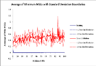
Fig.2
It is observed that MLP NN with one hidden layer with the 136-89-7-39 configuration yields the best results.
From the above experimentation, selected parameters for designing optimum MLP NN classifier are given below:
No. of inputs = 136, No. of hidden layers=01, No. of output PEs = 7, No. of epochs=1000,
For one hidden layer PEs and output layer PEs, for trans- fer function Tanh, Learning Rule Momentum NNhas been tested for training and testing the network.
For reliable clssification of lung images into three different types, classifier based on MLP NN have been developed and studied to get various variable parameters for optimum per- formance on Testing as well as cross-validation dataset.
The obtained Test Results are as shown belows: TABLE 2
Perfor- mance | Output(B) | Output(NL) | Output(M) |
MSE | 0.138479 267 | 0.261424548 | 0.186867629 |
NMSE | 0.553917 07 | 1.394264254 | 0.996627356 |
MAE | 0.343045 626 | 0.411481493 | 0.40802023 |
Min Abs Error | 0.155631 427 | 0.175869294 | 0.24379757 |
Max Abs Error | 0.510060 686 | 0.932950742 | 0.619114861 |
r | 0.748759 704 | -0.887960798 | 0.431000767 |
Percent Correct | 50 | 0 | 100 |
The proposed classifier is noticed as 100% on Tested data set .
The authors wish to thank Dr.R.S.Arora , Dr.S.V.Dudul , Prof.Vijay Dhawale, Dr.Varsha Agrawal to teach the Human Anatomy and basic process to use NeuroSoultion software and to Prof.R.N.Vaidya for type setting the paper.
[1] IEEE Transactions on Medical Imaging (April 1996, Vol. 15 , No.2) : Jyh-Shyan
Lin
[2] IEEE transaction on Medical Imaging(Dec.1998 , Vol.17,No.6):Manual
G.Penedo
[3] American Roentgen Ray Society (March 2002 ,AJR : 178)
: Yuichi Matsuki
[4] CHEST journal (January 2003): Bethany B.Tan,Kevin R.Flherty
[5] American Association for Artificial Intelligence(Flair-2003): C.F.Aliferis0
[6] Application of a Neural Network to Improve Nodal Staging Accuracy with F- FDGPET in Non-Small Cell Lung cancer(2003 ; 44:1918-1926): Hubert Vesselle
[7] Radiology(Nov.2005): Kwang Gi.Kim
[8] The Journal of Nuclear Medicine (2006 ; 47:1075-1080): Yongkang Nie
[9] Proceedings of the WSEAS International Conference on Signal, Speech and
Image processing Lisbon, Portugal ,(September 22-24 ,2006): Kim Le
[10] Medical Physics(March 2007,34(3)): Peng Wang
[11] IEEE/ICME: JIA TONG
[12] Cancer Biomarkers, IOC Press(2007): Michael Phillips,Nasser Altorki
[13] International Journal of Computer Applications,(2010,Vol.1- No.4):K.Balachandran, Dr.R.Anithha
[14] International Journal of Bio-science and Bio-Technology(June 2010 , Vol.2 , No.2) :Rishi Pal, Pradeep Garg
[15] IACIST International Journal of Engg. & technology (June 2010 , Vol.2, No.3)
: M.Gomathi and Dr.P.Thangaraj
[16] Digital Image Processing ,second edition ,PHI publication: Rafael C. Gonzalez
IJSER © 2013 http://www.ijser.org
International Journal of Scientific & Engineering Research, Vo lume 4, Issue 6, June-2013
ISSN 2229-5518
115
[17]Digital Signal Processing -Prindples ,Algorithms , and ApplkatiOllS, Fourth edition ,Pearson Education: Jolm G.Proakis, Dimitris G. manolakis
[18]New Atlas of Human Anatomy ,Lustre Press PvtLtd. :Thomas McCracken, GeneralEditor
[19] http:/ /www.wrongdiagnosis.com/c/cancer/introhtm?ktrack=kcphnk
IJSER lb)2013