International Journal of Scientific & Engineering Research, Volume 6, Issue 5, May-2015 791
ISSN 2229-5518
Evaluation of Monocyte Chemoattractant Protein-1 (MCP-1) in Type 2 Diabetes Mellitus.
Huda Jaber Waheed *,Ph.D, Muna Khalil**, MSc., Shahad Fawzi***, MSc.
*, ** University of Al-Mustansiriyah, College of Pharmacy, Clinical Laboratory Science Department, Iraq.
***Al-Israa University College, Department of Medical Lab Sciences, Iraq.
Abstract: AIM: To investigate the association of serum the chemokine monocyte chemoattractant protein-1 (MCP-1) levels, a major chemoattractant of monocytes and activated lymphocytes, with metabolic parameters like insulin hormone, glycolated hemoglobin and glucose Iraqi patients with type 2 diabetes mellitus.
METHODS: MCP-1 , TGF-β1 and insulin concentrations were measured by enzyme-linked immunosorbent assay (ELISA), while
Hba1c and glucose were measured by spectrophotometer technique.
RESULTS: Thirty diabetic patients were compared with 20 healthy subjects to assess the studied parameter. MCP-1 mean concentrations and percentiles were substantially higher in non-diabetic populations. MCP-1 serum levels are related to age, BMI, HbA1c, TGF- β1, glucose and insulin hormones
CONCLUSIONS: Compared to healthy groups, MCP-1 levels were found to be substantially higher in patients with type 2 diabetes mellitus.
Keywords: MCP-1, hyperglycemia, diabetes mellitus, TGf-β1.
1. Introduction
—————————— ——————————
Diabetes mellitus is considered one of the most vicious chronic diseases of our time, owing to the fact that, individuals have to endure long years of successive complications, makes it less probable to be controlled. Diabetes mellitus is a metabolic disorder identified by a lingering hyperglycemia as a result of defect in insulin secretion, insulin action or both. Which impairs the body's ability to metabolize sugar, lipid and protein. The effects of diabetes include long-term damage, dysfunction and failure of different organs[1]. Type2 diabetes comprises 95% of diabetic cases worldwide and its etiology is most probably related obesity and insulin resistance [2].
Chemokines (chemotactic cytokines) are small heparin-binding proteins which direct migration of circulating leukocytes to
sites of inflammation or injury [3]. The largest family is the CC chemokine MCP-1( monocyte-chemo-attractant protein 1). Two residues of MCP 1, out of four conserved cysteine residues are thoroughly characterized CC chemokine. MCP-1 (monocyte chemo-attractant protein 1), also known as CCL2 – a potent agonist for monocytes, memory T cells and basophils intimal hyperplasia after angioplasty, as well as in vas-culogenesis and thrombosis [4]. MCP-1 is also expressed and secreted by adipocytes, which has been reported to be involved in the recruitment and activation of peripheral blood leukocytes in adipose tissue and in the induction of systemic insulin resistance [5]. Another study has reported the identification of a novel protein; designated MCPIP (MCP-induced protein), which is expressed in human monocytes and cardio- myocytes after stimulation with MCP-1.
IJSER © 2015 http://www.ijser.org
International Journal of Scientific & Engineering Research, Volume 6, Issue 5, May-2015 792
ISSN 2229-5518
Their results provided a novel molecular pathway, by which MCP-1 signal transduction is linked to transcriptional gene regulation leading to apoptosis [6].
Hyperglycemia is the major cause of diabetic angiopathy. High glucose treatment on endothelial cells isolated from diabetic subjects resulted in a 40-70% increase of MCP-1 release, and a 10-20% increase of the basal expression of vascular cell adhesion molecule-1 (VCAM-1), indicating synergistic enhancement on the monocyte- endothelial cell interaction [7]. In a vitro study demonstrated that advanced glycation end-products; high glucose concentration, glycated albumin and glycoxidized LDL enhanced MCP-1 expression in human endothelial cells [8]. Similarly, high glucose treatment on human aortic smooth muscle cells (SMC) unregulated the expression of MCP-1 and fractalkine leading to increased monocyte-SMC adhesive interactions by a
TGF ((DRG diagnostics Company, Germany). insulin (Demedetec Company, Germany) were detected by enzyme-linked immunosorbent (ELISA).
2.1 Statistical analysis
Analysis of data was carried out using the available statistical package of SPSS-22 (Statistical Packages for Social Sciences- version 22). The significance of difference of different means (quantitative data) were tested using Students-t-test or ANOVA test. The significance of difference of different percentages (qualitative data) were tested using Pearson Chi-square test (χ2-test) with application of Yate's correction or Fisher Exact test whenever applicable. Statistical significance was considered whenever the P value for the test of significance was equal or less than 0.05.
3. Results:
mechanism involving activation of MAPK, AP-1 and NFκB [9] . Consistent with previous reports, exposure of human endothelial ECV304 cells to high glucose for
24 h caused an increase of MCP-1 and intercellular adhesion molecule-1 (ICAM-1), and promoted cell adhesion between
monocyte and ECV304 cells[10]. Furthermore, high glucose treatment on human acute monocytic leukemia THP-1 cells increases both mRNA and protein levels of MCP-1, enhanced the adhesion of THP-1 cells to endothelial cells, and the pathways reportedly involved oxidative stress and protein kinase C [11].
The present study is aimed to assess the levels of MCP-1 and other biochemical markers in diabetic subjects compared to a healthy group. Which in turn can be beneficial to clarify the correlations of MCP-
1 levels in diabetes mellitus with other clinical parameters.
2. Subject and Methods:
Twenty normal subjects (aged 53.33 ±


6.195 years; mean SD) and 30 diabetic patients were studied (aged 56.27 ± 5.587 years; mean SD). Informed consent was
obtained from each subject. Patients with concurrent acute illnesses, malignancy, and active immunological diseases; medical history of clinical cardiovascular disease; medical history included the diseases (hypertension, rheumatoid arthritis, anemia, bronchial asthma) or medications (warfarin, acetylsalicylic acid, alpha-methyldopa, vitamins, tramadol,simvastatin) that interfered with HbA1c % measurements, and smoking history were excluded from the study. The anthropometric measurements and blood pressure were recorded. Biochemical testing included plasma fasting blood glucose (LABKIT,Spain), Glycated hemoglobin HbA1c % (Human Co., German), MCP-
1( Elabscience Biotechnology Co. Japan),
IJSER © 2015 http://www.ijser.org
International Journal of Scientific & Engineering Research, Volume 6, Issue 5, May-2015 793
ISSN 2229-5518
patient and healthy subject, as shown in the table(1) . Table (1) also shown the duration of diabetes in the patients also recorded in years.
Patients and control group were similar in regard to age with no significant difference [P value >0.05] , as well as there were no significant difference between diabetic
Table (1): Means of Age, BMI and duration of disease in T2DM patients and controls.
Parameters (mean ± SD) | Controls (n = 20) | Patients (n = 30) | P.value |
Age | 48.40±6.53 | 51.29±7.62 | 0.48 |
BMI | 29.53 ± 1.552 | 29.93 ± 1.792 | 0.45 |
Duration of disease (year) | 4.46±4.69 | - | - |
Table (2) show the core study analytes. Serum MCP-1 level was significantly higher in diabetic patients than
in control subjects (48.87 ± 12.345 vs. 38.8 ±
8.994 ng/ml respectively, p<0.001), figure
(1).
Table (2):- Comparison between biochemical parameters of controls and patients subjects.
Parameters (mean ± SD) | Controls (N =20) | Patients (N = 30) | P.value |
MCP1 (ng/ml) | 38.8 ± 8.994 | 48.87 ± 12.345 | 0.003** |
TGF-β1 (pg/ml) | 13.6 ± 4.641 | 19.67 ± 6.586 | 0.03 |
Glucose (mg/dl) | 83.8 ± 9.689 | 130.8 ± 16.575 | < 0.001 * |
Insulin (µIU/ml) | 20.93 ± 4.758 | 41.13 ± 8.132 | < 0.001 * |
HbA1c | 4.68 ± 0.4843 | 7.993 ± 0.6464 | < 0.001 * |
* The P-value ≤ 0.001 at degree of freedom 14 is highly significant.
** The P-value ≤ 0.05 at degree of freedom 14 is significant.
IJSER © 2015 http://www.ijser.org
International Journal of Scientific & Engineering Research, Volume 6, Issue 5, May-2015 794
ISSN 2229-5518
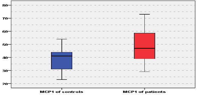
Figure (2):- The Boxplot graph of MCP1 of controls and patients.
The Boxplot graph (2) shows the difference between the means of TGF of controls and patients. The T2DM patients showed a slightly increased mean of TGF in
comparison with controls (19.67 ± 6.586 vs.
13.6 ± 4.641pg/ml) and the difference was not significant.
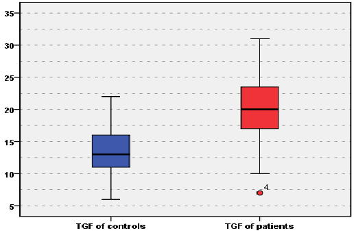
Figure (3):- The Boxplot graph of TGF of controls and patients.
The mean of insulin serum level was highly significantly increased in diabetic patients groups (41.13 ± 8.132 µIU/ml ) as compared to controls (20.93 ± 4.758 µIU/ml)
IJSER © 2015 http://www.ijser.org
International Journal of Scientific & Engineering Research, Volume 6, Issue 5, May-2015 795
ISSN 2229-5518
. There was a significant difference (P<0.001) in both Hba1c and plasma glucose between studied groups (using t- test ), table (2).
In diabetic patients, there were positive highly significant correlation between MCP-1 and age, BMI, HbA1c , TGF- β1, glucose and insulin ( r = 0.359, r = 0.717,
r = 0.395, r = 0.217, r = 0.595 and r = 0.558 respectively, P <0.01), as well as TGF- β1 also significantly correlated with BMI, HbA1c, MCP-1, glucose and insulin( r =
0.606, r = 0.871, r = 0.595, r = 0.217, r =
0.595, r = 0.367 and r=0.178 respectively), while both MCP-I and TGF- β1were
negatively correlated with age ( r = -0.359 and r = -0.178 respectively), table (3).
Table (3):- Correlation between MCP-1 and TGF- β1 with other biochemical parameters of patient subject.
| | Age | BMI | HbAR1Rc | MCPR1 | TGF | FPG | Insulin |
| N | 30 | 30 | 30 | 30 | 30 | 30 | 30 |
MCPR1 | Pearson Correlation | -0.359* | 0.717*** | 0.395* | --- | 0.217* | 0.595** | 0.558** |
MCPR1 | P.value | 0.189 | 0.003 | 0.145 | --- | 0.437 | 0.019 | 0.031 |
TGF- β1 | Pearson Correlation | -0.178* | 0.606** | 0.871** * | 0.595** | --- | 0.367* | 0.774*** |
TGF- β1 | P.value | 0.526 | 0.017 | <0.001 | 0.019 | --- | 0.178 | 0.001 |
4. Discussion
MCP-1 is a potent chemo-attractant for monocytes, after a thorough research has been conducted and successive measurements have been taken, the study showed that; there is a significant increment in diabetic subjects when compared to a healthy group. These findings are corresponded to the well known metabolic changes that occur in normal and diabetic subjects. Healthy individuals (non-diabetic) produce an "accelerated starvation" in the fasting state, with an earlier and more
profound hypoglycemia and an increased fasting insulin level, whereas diabetic patients; first of all, demonstrate an elevated fasting insulin concentrations [4]. Nelson et al. With a significant release of MCP-1 from peripheral blood cultures from pregnant women as compared to non-pregnant women[12].
Moreover, study has shown that; there were a significant positive correlation between MPC-1 and BMI , MCP-1 signaling which has a direct role in the development of obesity. For example, Zue CW et al. has
IJSER © 2015 http://www.ijser.org
International Journal of Scientific & Engineering Research, Volume 6, Issue 5, May-2015 796
ISSN 2229-5518
reproted that, MCP-1-induced protein (MCPIP, a zinc finger protein) induced adipogenesis in 3T3-L1 cells independent of PPARgamma activation [13 ]. Moreover, MCP-1 had angiogenic effect on endothelial cells , and therefore it can contribute to the expansion and remodeling of adipose tissues [14] [15].
One of the studied factors was; transforming growth factor - beta1 (TGF-beta1) - a multifunctional cytokine that exhibits potent immunoregulatory and anti-inflammatory properties and prolongs graft survival [16].
Beata Telejko hypothesize that TGF-beta may be a key factor responsible for the alterations in circulating MCP-1 levels, but the secretion and action of this chemokine in diabetes mellitus need further investigations[4].
In conclusion, the current study's findings
suggest that; glycemic status influences serum MCP-1 levels in diabetic patients, where MCP-1 strongly is correlated with glycemic parameter ( insulin hormone, Hba1c, glucose) . Elevated serum MCP-1 levels could contribute to the onset and progression of several complications in diabetes. Thus, serum MCP-1 may serve as a biomarker of inflammatory activity and helps in early detection and intervention of diabetic complications.
ACKNOWLEDGMENT
We are thankful to all staff of National
Diabetic Center, Al-Mustansiriyah
University for providing us facilities and kind support throughout the research work.
REFERENCE
1. World Health Organization.
Definition, Diagnosis and
Classification of Diabetes Mellitus
and its Complications. Second Report. Geneva: WHO, 1999. Technical Report Series646.
2. Piemonti L, Calori G, Lattuada G, Mercalli A, Ragogna F, Garancini MP, et al. Association between plasma monocyte chemoattractant protein-1 concentration and cardiovascular disease mortality in middle-aged diabetic and nondiabetic individuals. Diabetes Care.
2009;32:2105–10.
3. Charo IF, Taubman MB. Chemokines
in the pahogenesis of vascular disease.
Circ Res. 2004;95:858-866.
4. Beata Telejko, Mariusz Kuzmicki, Anna Zonenberg, et al. Circulating
monocyte chemoattractant protein-1 in women with gestational diabetes. FOLIA Histochemica ET Cytobiologica Vol. 45, Supp. 1, 2007 pp. 153-156]
5. Zaica RE. MCP-1 (monocyte chemotactic protein-1) and harpin
molecules in inflammation processes .
Bio Med, 2014, 44(7);213-217.
6. Zhou L, Azfer A, Niu J, Graham S,
Choudhury M, Adamski FM, Younce C, Binkley PF, Kolattukudy PE. Monocyte chemoattractant protein-1 induces a novel transcription factor that causes cardiac myocyte apoptosis and ventricular dys- function.Circ Res.2006;98:1177-1185.
7. Jun Panee. Monocyte Chemoattractant Protein 1 (MCP-1) in Obesity and Diabetes. Cytokine. 2012
Oct; 60(1): 1–12.
8. Ihm CG, Park JK, Hong SP, Lee TW, Cho BS, Kim MJ, Cha DR, Ha H. A high glucose concentration stimulates the expression of monocyte chemotactic peptide 1 in human mesangial cells. Nephron.1998;79:33-
37.
9. Dragomir E, Manduteanu I, Calin M, Gan AM, Stan D, Koenen RR, et al. High glucose conditions induce
IJSER © 2015 http://www.ijser.org
International Journal of Scientific & Engineering Research, Volume 6, Issue 5, May-2015 797
ISSN 2229-5518
upregulation of fractalkine and monocyte chemotactic protein-1 in human smooth muscle cells. Thromb Haemost. 2008;100:1155–65
10. Luo P, Tan ZH, Zhang ZF, Zhang H, Liu XF, Mo ZJ. Scutellarin isolated from Erigeron multiradiatus inhibits
high glucose-mediated vascular inflammation. Yakugaku Zasshi.
2008;128:1293–9
11. Shanmugam N, Reddy MA, Guha M, Natarajan R. High glucose-induced expression of proinflammatory cytokine and chemokine genes in monocytic cells. Diabetes.
2003;52:1256–64.
12. Nelison B.R, Katiz RW, Anna AA.
Differential secretion of chemokines from peripheral blood in pregnant women compared with non - pregnant women. Australian Journal of diabetes. 2014;34:125-140
13. Zue CW, Mohoto A. MCP-1 (monocyte chemotactic protein-1)- induced protein, a recently identified zinc finger protein. J Biol Chem.
2011;284:27620–8.
14. Younce M., Nasif T., Redhah A.
Assessment of MCP-1 and VEGF in
a study case, Endo. Bio Jou, 2014 ;
12(7); 311-313.
15. kado R et al. Human endothelial cells
express CCR2 and respond to MCP-1: direct role of MCP-1 in angiogenesis and tumor progression. Bio. Chem.
2010;96:34–40
16. Hong Xie. Effect of the cytokines on successful pregnancy depends upon the control of graft rejection mechanisms. Transplant Journal.
2014;39:174-175
IJSER © 2015 http://www.ijser.org