The research paper published by IJSER journal is about A new masks group called A. H. SH. Rostom for Mycosis Fungoides Skin image Edge detection 1
ISSN 2229-5518
A new masks group called A. H. SH. Rostom for
Mycosis Fungoides Skin image Edge detection
Dr. Ali Hassan Nasser Al-Fayadh Assistant .Professor. Mathematic Department Mathematical and Computer Sciences College Kufa University, Iraq | Hind Rostom Mohammed Assistant Professor Computer Department Mathematical and Computer Sciences College/ Kufa University, Iraq | Shaymaa Maki kadham Mathematic Department Mathematical and Computer Sciences College Kufa University, Iraq |
aalfayadh@yahoo.com | hindrustum.shaaban@uokufa.edu.iq | a.alkhafajee@yahoo.com |
Abstract—In the present paper, new method called (A.H. SH..Rostom Group masks) for Mycosis Fungoides Skin image Edge detection is proposed. The Group consists of 10 masks were geometry of the mask operator determines a characteristic direction in which i t is most sensitive to edges. applied to the four stages of the Mycosis Fungoides disease Skin image have been identified and the edges of the images used for each and every stages that the database consists of 40 images divided each stage of the Mycosis Fungoides dis ease Skin image 10 images. For each stage a novel algorithm which combines pixel and regi on based color segmentation techniques is used. The experimental results confirm the effectiveness of the proposed A.H. SH.Rostom Group masks .
Index Terms—Edge detection ,Skin image detection, Segmentation, Image processing.
—————————— ——————————
1 INTRODUCTION
Edge detection is an important field in image processing. It can be used in many applications such as segmentation,
registration, feature extraction, and identification of objects in a scene. An effective edge detector reduces a large amount of data but still keeps most of the important feature of the image. Edge detection refers to the process of locating sharp disconti- nuities in an image. These discontinuities originate from dif- ferent scene features such as discontinuities in depth, discon- tinuities in surface orientation, and changes in material prop- erties and variations in scene illumination[1,2].
The boundaries of object surfaces in a scene often lead to oriented localized changes in intensity of an image, called edges. This observation combined with a commonly held be- lief that edge detection is the first step in image segmentation, has fueled a long search for a good edge detection algorithm to use in image processing [3]. Edge detection of an image re- duces significantly the amount of data and filters out informa- tion that may be regarded as less relevant, preserving the im- portant structural properties of an image. Therefore, edges detected from its original image contain major information, which only needs a small amount of memory to store[4].
Edge detection produces something like a line drawing of an
image, which highlights the intensity changes. In general, the
boundaries of objects tend to produce sudden changes in the
image intensity[5]. different surfaces of an object receive dif-
ferent amounts of light, which again produces intensity
changes[6]. Edges are effected by noise present in an image
though .An edge may be regarded as boundary between two
dissimilar regions in an image .edge detection is a terminology
in image processing and computer vision , particularly in
areas of feature detection and feature extraction[7].
The paper is organized as follows; Section 2 deals with the
A.H. SH.Rostom Group masks are considered to determine
the Mycosis Fungoides Skin image area. Section 3 deals with the Edge Operations i.e opening to perform the connected component analysis, section 4 gives the overview of algorithm with Experimental Results and last section 5 ends the paper with conclusion
2 A.H. SH.ROSTOM GROUP MASKS
A human skin color model is used to decide either a pixel is skin color or non skin-color[5]. In this research, we use mew method called(A.H. SH.Rostom Group masks ) Mycosis Fun- goides Skin image edge detection . A.H. SH Rostom Group masks consists of 10 masks determines a characteristic direc- tion edge.
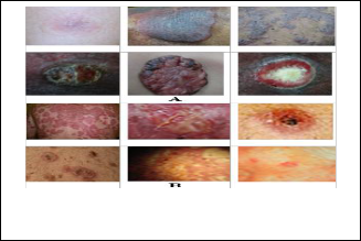
The Details of values for each mask in A.H. SH.Rostom Group masks shown in figure(1) and the skin images library sam- ples, two types of images (A)Samples with Mycosis Fungoides diseases Skin images (B) Samples with other diseases shown in figure(2).
M1 M2 M3
M4 M5 M6
M7 M8 M9
IJSER © 2012
http://www.ijser.org
The research paper published by IJSER journal is about A new masks group called A. H. SH. Rostom for Mycosis Fungoides Skin image Edge detection 2
ISSN 2229-5518

M10
Figure(1): A.H. SH.Rostom Group masks
A.H. SH.Rostom Group masks determined for direction edge:
M1= Vertical1 ,M2=Horizontal1 M3= Vertical-Horizontal1
M4= Vertical2 M5= Horizontal2 M6= Vertical-Horizontal2
M7=Diagonal1 , M8= Diagonal2 M9= Diagonal1- Diagonal2
M10=Mask roots
Figure(2): The skin library samples, (A) Samples with Skin dis- eases (B) Samples with other diseases
The skin is involved, in addition to either of the following:
• Cancer cells are found in the lymph nodes;
• Cancer has spread to other organs, such as the liver or lung[8].
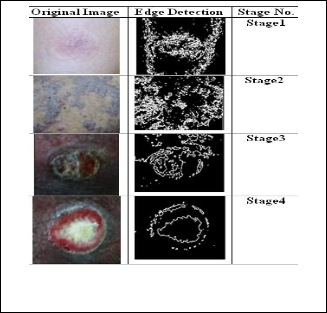
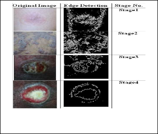
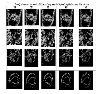
Experimental Results for Appling Edge Detection mask shown in figure (3,4,5,6,7,8,9,10,11,12).
4 CONCLUSION
new method called (A.H. SH.Rostom Group masks) for Myco- sis Fungoides Skin image edge detector presented in this pa- per uses ten masks determines a characteristic direction in which it is most sensitive to edges . The proposed method is decrease the computation time with generate high quality of edge detection. Experiment results have demonstrated that the proposed scheme for edge detection works satisfactorily for different levels digital images. Another benefit comes from easy implementation of this method. A.H. SH.Rostom Group masks for Mycosis Fungoides Skin image Edge detection is necessary to provide a robust solution that is adaptable to the varying noise levels of these images to help distinguish valid image contents from visual artifacts introduced by noise. The experimental results show the satisfying subjective test results and The simulation results are very promising.
3 EXPERIMENTAL RESULTS
In this section a detailed experimental comparison of the above stated A.H. SH.Rostom Group masks has been presented. We have used two types Mycosis Fungoides Skin image databases:
(1) database prepared in our conditions ,images obtained from in
Al-Sder Hospital.
(2) Skin database [4] and some other images obtained from in- ternet.
Mycosis fungoides is a T-cell lymphoma of the skin. The disease is caused by the proliferation of T-lymphocytes, also known as helper T cells[8].
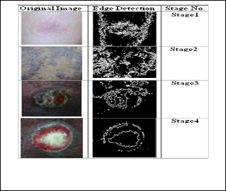
In this paper divided stages images as
Stages in mycosis fungoides(10 images for each stage) Stage 1
The cancer only affects parts of the skin, which has red, dry, scaly
patches, but no
tumours. The lymph nodes are not larger than normal. Stage 2
Either of the following may be true:
• The skin has red, dry, scaly patches, but no tumours. Lymph
nodes are larger than
normal, but do not contain cancer cells;
• There are tumours on the skin. The lymph nodes are either
normal or are larger than
normal, but do not contain cancer cells. Stage 3
• Nearly all of the skin is red, dry, and scaly. The lymph nodes are either normal or are larger than normal, but do not contain can- cer cells.
Stage 4
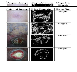
Figure(3):(A):Original Skin Image , (B) Edge detection by M1 for
A for all Mycosis Fungoides Skin image stages
IJSER © 2
http://www.ijs
The research paper published by IJSER journal is about A new masks group called A. H. SH. Rostom for Mycosis Fungoides Skin image Edge detection 3
ISSN 2229-5518
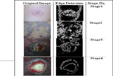
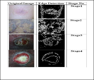
Figure(5):(A):Original Skin Image , (B) Edge detection by M3 for
A for all Mycosis Fungoides Skin image stages
Figure(8):(A):Original Skin Image , (B) Edge detection by M3 for
A for all Mycosis Fungoides Skin image stages
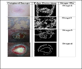
Figure(6):(A):Original Skin Image , (B) Edge detection by M3 for
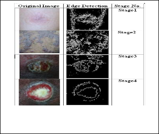
A for all Mycosis Fungoides Skin image stages Figure(9):(A):Original Skin Image , (B) Edge detection by M3 for
A for all Mycosis Fungoides Skin image stages
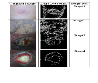
Figure(7):(A):Original Skin Image , (B) Edge detection by M3 for
A for all Mycosis Fungoides Skin image stages
Figure(10):(A):Original Skin Image , (B) Edge detection by M3 for A for all Mycosis Fungoides Skin image stages
IJSER © 2012
http://www.ijser.org
The research paper published by IJSER journal is about A new masks group called A. H. SH. Rostom for Mycosis Fungoides Skin image Edge detection 4
ISSN 2229-5518
[9] Steven M. Horwitz, MD;a Elise A. Olsen, MD;b Madeleine Duvic, Review of the Treatment of Mycosis Fungoides
[10] and Sezary Syndrome: A Stage-Based Approach, Journal of the Na- tional Comprehensive Cancer Network Volume 6 Number 4 April
2008.
.
Figure(12):(A):Original Skin Image , (B) Edge detection by M3
for A for all Mycosis Fungoides Skin image stages
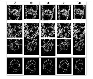
Table (1) shown the comparison for A.H. SH Rostom Group masks for Mycosis Fungoides Skin image Edge detection. Edge detection are computationally more expensive compared to M1, M2, M4, M5, M7 and M8 masks. However, the M10 edge detection algorithm performs better than all these opera- tors under almost all scenarios. Evaluation of the images showed that under noisy conditions, M10, M9, M6 and M3 exhibit better performance, respectively.
REFERENCES
[1] Mohamed A. El-Sayed, A New Algorithm Based Entropic Thre- shold for Edge Detection in Images, IJCSI International Journal of Computer Science Issues, Vol. 8, Issue 5, No 1, September 2011.
[2] A. El-Zaart, "A Novel Method for Edge Detection Using 2 Dime n- sional Gamma Distribution", Journal of Computer Science 6 (2), 2010.
[3] N. Senthilkumaran and R. Rajesh, “A Study on Edge Detection Me- thods for Image Segmentation”, Proceedings of the International Conference on Mathematics and Computer Science (ICMCS-2009), Vol.I 2009.
[4] C.NagaRaju , S.NagaMani, G.rakesh Prasad, S.Sunitha., Morphologi-
cal Edge Detection Algorithm Based on Multi-Structure Elements of
Different Directions,
[5] International Journal of Information and Communication Technolo- gy Research, Volume 1 No. 1, May 2011.
[6] Balkrishan Ahirwal, Mahesh Khadtare and Rakesh Mehta, “FPGA based system for Color Space Transformation RGB to YIQ and YCbCr.” International Conference on Intelligent and Advanced Sys- tems,2007.
[7] B.Poornima, Y.Ramadevi, T.Sridevi, Threshold Based Edge Detec- tion Algorithm, IACSIT International Journal of Engineering and Technology, Vol.3, No.4, August ,2011.
[8] Beant Kaur, Anil Garg, Amandeep Kaur, Mathematical Morpholog- ical Edge Detection For Remote Sensing Images, IJECT Vol. 1, Issue 1,
December 2010.
IJSER © 2012
http://www.ijser.org