Of the 200 subjects studied the commonest sonologic marker observed was pyelectasis (n=6) followed by choroid plexus cyst (n=5).
International Journal of Scientific & Engineering Research, Volume 5, Issue 7, July-2014 1126
ISSN 2229-5518
Validity of Sonologic Soft Marker for Chromosome
Abnormality
Laly Jose, Alex K Ittyavirah and Dinesh Roy
—————————— ——————————
Chromosomal abnormalities are one of the leading causes of pregnancy loss. Chromosome abnormalities occur in 0.1-0.2% of all live births [1]. Trisomy 21 is the most common karyotypic abnormality in live born infants [2]. Sonographic finding in fetuses with Down syndrome include both structural and non-structural abnormalities. Second trimester ultrasound detects two types of sonographic findings suggestive of aneuploidy. Detection of major fetal structural anomalies comprises the first group. The second group includes soft markers that are non specific often transient and can be detected during 2nd trimester ultrasound scan [3]. The most commonly studied soft markers of aneuploidy included nuchal fold thickness, rhizomelic limb shortening, mild pyelectasis, echogenic bowel, echogenic intra cardiac focus and choroid plexus cyst. Unfortunately the studies evaluating significance of soft markers for aneuploidies vary widely and show contradictory results. The present study reviews the validity of sonographic markers for chromosome abnormalities in study population to identify the best marker for detecting at risk pregnancy for chromosome abnormality.
————————————————
• Laly Jose, Associate Professor of Radiodiagnosis, SM CSI Medical
College, Karakonam P.O, Thiruvananthapuram- 695504.
• Alex K Ittyavirah and Dinesh Roy, Ittyavirah Scans and Genetic
Research, Thiruvananthapuram- 695011.
The study was conducted on 200 subjects referred from Obstetrics and Gynaecology OPD of SM CSI Medical College, Karakonam, Trivandrum for a period of 2 years.
Inclusion criteria:
1. Pregnant subjects below 35 years of age.
2. Subjects without any added risk for chromosome
abnormality.
The study was done using Siemens sonolin 50 USS
scanner. The following soft markers were evaluated during
18-20wks scan.
Nuchal fold thickness
The measurement is made in transverse plane of fetal head slightly off the biparietal diameter which includes cerebellum, occipital bone and cavum septum pellucidum. The Nuchal fold was measured with placement of calipers from outer edge of occipital bone to outer edge of skin [4]. 5mm was used as single cut off.
Echogenic bowel
Fetal echogenic bowel refers to presence of hypoechoic bowel as compared with echogenicity of adjacent iliac bone. Once echogenic bowel was suspected the gain of USS unit was lowered gradually until only bone or bowel was visible.
Short long bones
Shortened humerus or femur is identified by comparing actual measurement with expected measurement on the basis of biparietal diameter. The femur is considered shortened when measured to expected ratio that is < 0.89 [5].
IJSER © 2014 http://www.ijser.org
International Journal of Scientific & Engineering Research, Volume 5, Issue 7, July-2014 1127
ISSN 2229-5518
Echogenic Intracardiac focus
Echogenic Intracardiac focus is seen as discrete areas of echogenicity compared to bone in the region of papillary muscle. The foci were visualized from different angles to make sure that one does not include specular reflections of papillary muscle.
Choroid plexus cyst
Looked for in axial plane of head within lateral ventricle. Mild pyelectasis
Mild pyelectasis is diagnosed when renal pelvis measures > 4 mm and
<10 mm in AP dimension in axial scan of abdomen without caliceal dilatation.
Karyotyping
Karyotyping was done in case of subjects with positive soft markers for chromosome abnormality using the blood collected by cordocentesis of foetus or by cord blood of baby. Karyotyping was done by lymphocyte culture method at Ittyavirah Scan and Genetic Research.
Of the 200 subjects studied the commonest sonologic marker observed was pyelectasis (n=6) followed by choroid plexus cyst (n=5).
IJSER © 2014 http://www.ijser.org
International Journal of Scientific & Engineering Research, Volume 5, Issue 7, July-2014 1128
ISSN 2229-5518
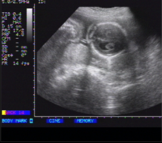
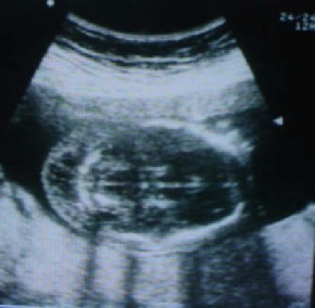
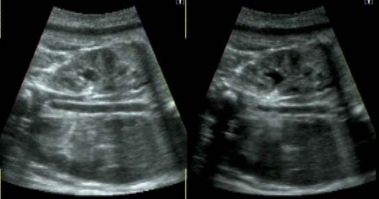
FIG. 3 (a &b) Minor pyelectasis
Sl. | Soft marker | Number | Percentage |
1 | Nuchal thickness | 2 | 1 |
2 | Choroid plexus cyst | 5 | 2.5 |
3 | Echogenic bowel | 1 | 0.5 |
4 | Pyelectasis | 6 | 3 |
5 | Short long bone | 1 | 0.5 |
6 | Echogenic Intracardiac focus | 4 | 2 |
Total | 19 | 9.5 |
TABLE 1:
Sonographic markers observed in 200 antenatal patients
IJSER © 2014 http://www.ijser.org
International Journal of Scientific & Engineering Research, Volume 5, Issue 7, July-2014 1129
ISSN 2229-5518
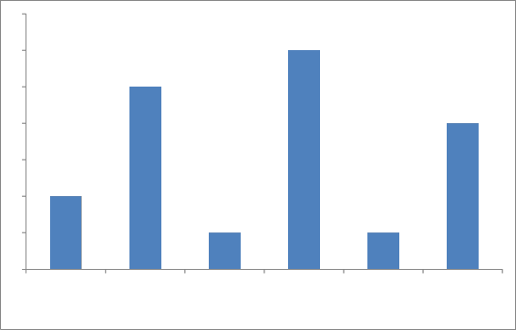
7
6
6
5
5
4
4
3
2
2
1 1
1
0
Nuchal thickness
Choroid plexus cyst
Echogenic bowel
Pyelectasis Short long bone Echogenic
Intracardiac focus
FIG. 4
Sonographic markers observed in 200 antenatal patients
Soft markers | Number | Abnormal karyotype | |
1 | Choroid plexus cyst and Nuchal thickness | 6 | 0 |
2 | Echogenic bowel and short long bone | 1 | 1 |
3 | Choroid plexus cyst with echogenic bowel | 2 | 1 |
4 | Pyelectasis and NT | 4 | 0 |
Total | 13 | 2 |
TABLE 2:
Multiple sonographic markers in 200 antenatal patients and their correlation with chromosome abnormality
IJSER © 2014 http://www.ijser.org
International Journal of Scientific & Engineering Research, Volume 5, Issue 7, July-2014 1130
ISSN 2229-5518
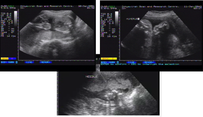
FIG. 7
Cordocentesis under ultrasound guidance
Out of the 200 subjects included in the present
study 19 subjects had solitary sonographic soft marker for chromosome abnormality. The commonest marker observed was pyelectasis followed by choroid plexus cyst. None of the cases with solitary soft marker had chromosome abnormality detected by karyotyping. Among the 200 subjects 9 subjects had multiple soft markers; out of these, 2 fetuses had chromosome abnormality. One was Down syndrome and the other was Turner’s syndrome.
In the present study out of 200 subjects, 19 antenatal cases showed isolated soft marker for chromosome abnormality and none of these fetuses had chromosome abnormality on karyotyping. Nyberg DA et al reported that isolated soft marker was the only sonographic finding in 42 (22.6%) of 166 fetuses with trisomy. When compared with 11% of control population [6], in this study the percentage of isolated marker in subjects is 9.5%. According to Bomle B et al [7] echogenic intracardiac focus was the single most common isolated marker in both affected (7.1%) and control fetus (3.9%). In this study multiple marker was noted in 13 antenatal cases which included 6 cases of choroid plexus cyst with NT, 4 cases of
pyelectasis with NT, 2 cases of choroid plexus cyst with
echogenic bowel and one case of echogenic bowel with short long bone. Out of the 9 cases with multiple markers, 2 fetuses had chromosome abnormality. Out of the 19 subjects those showed solitary marker none of them had chromosome abnormality which suggesting that cluster of markers seen to confer higher risk of aneuploidy than solitary marker. Similar conclusions were made by Sohi B et al [8]. According to Sameer Ramga et al, Nuchal fold thickening, short humerous, even as isolated findings, confirm high risk of aneuploidy [9].
Out of 200 subjects included in the study 19 had solitary soft marker for chromosome abnormality and none of them had chromosome abnormality. Out of the 9 subjects with multiple soft markers, 2 fetuses had chromosome abnormality. There is no isolated soft marker is found to have higher risk for chromosome abnormality whereas cluster of markers seems to confer higher risk for aneuploidy.
IJSER © 2014 http://www.ijser.org
International Journal of Scientific & Engineering Research, Volume 5, Issue 7, July-2014 1131
ISSN 2229-5518
1. Shipp TD, Bena Ceraf BR. Second trimester ultrasound screening for chromosomal abnormalities. Prenat. Diag.
2002; 22: 296-307.
2. Thompson M, Mclnnes R, Willard H, Thompson and
Thompson, Genetics in Medicine. 5th edn. Philadelphia
WB. Saunder. 1001.
3. Vandenhrf MC, Watson RD. Fetal soft markers in obst.
USS – J. Obst. Gynae. 2005: 27 (6); 592-636.
4. Ship TD, Benaceraf BR. Second trimester USS screening
for chromosomal abnormalities – Pre nat. Diag. 2002; 22:
295-307.
5. Nyberg DA, Resta RG, Luthy DA, Hickok DE, William
MA. Humerus and femur length shortening in detection of Down syndrome. Am. J. Obst. Gynaecology.
168: 534-539.
6. Nyberg DA, Souter VL, Bastawiss A, Young S,
Luthhordt F, Luthy DA. Isolated sonographic markers
for detection of fetal down syndrome in 2nd trimester of pregnancy. J. Ultrasound Medicine. 2001: 20; 1053-1063.
7. Brimley B, Lieberman E, Shipp TD, Benacerraj BR.
Genetic sonogram a method of risk assessment for
down syndrome in 2nd trimester. Ultrasound. Med.
2002; 21; 1087-1096.
8. Sobi B, Sciascia A, Budorick NE, Moore TR. Utility of
minor sonographic markers in prediction of abnormal fetal karyotyping at a prenatal diagnostic centre. Am. J. Obst. Gynae. 1999; 181: 893-903.
9. Sameer Ramga. PD, Desai H, et al. Parikh Med. Scape; General Medicine. 2006; 8(1) : 9.
IJSER © 2014 http://www.ijser.org