International Journal of Scientific & Engineering Research, Volume 5, Issue 7, July-2014 587
ISSN 2229-5518
Synthesis and Characterization of Barium Titanate, Calcium Titanate and Strontium Titanate Thin Films
P.N.Nirmala*and G.Suresh
Department of Engineering Physics, Faculty of Engineering and Technology, Annamalai University, Chidambaram –
608002,Tamil Nadu,India.
*Corresponding author: pnnirmala@yahoo.com
Abstract: Barium titanate, Calcium titanate and Strontium titanate nano structured thin films have been successfully grown on glass substrate by Chemical Bath Deposition (CBD) technique. These films were characterized by scanning electron microscopy with energy dispersive analysis, X-ray diffraction and UV analysis. From the UV study the band gap of Barium titanate, Calcium titanate and Strontium titanate were 1.8eV, 2.17eV and 1.9eV.
Index Terms: Thin films, CBD, scanning electron microscopy, X-ray diffraction
—————————— ——————————
Thin films may be amorphous/nanocrystalline, polycrystalline and preferentially oriented or, very rarely, single crystal structures. Different structures have different properties. For example, in capacitor applications the crystalline structures generally have higher dielectric constants than the amorphous structures. This might be due to the increase in tetragonality of the BaTiO3. Preferentially oriented or textured thin films may be better suited for some applications compared to other structures. Since as- dried films are amorphous or very weakly crystalline, nucleation and growth processes determines the type of film structure that can be expected. Nucleation in the bulk of the film as well as the film-substrate interface usually leads to polycrystalline thin films. The remaining organics or precursors influence the nucleation of new crystals and the final structure of the films. For these films, the growth depends greatly upon nucleation at the film-substrate interface. The grains which grow from the interface gradually consume the small, randomly oriented grains in the film bulk.
Alkaline earth titanates MTiO3 (M = Ba, Ca and Sr ) with a cubic perovskite structure have been investigated intensively due to their unique dielectric, piezoelectric, and ferroelectric properties, which are of great interest in the technological applications such as capacitors, transducers, actuators, and nonvolatile random-access memory devices [1] . Moreover, SrTiO3 is an important n-type semiconductor with band gap of about 3.2 eV [2]. The stability, wavelength response, and current-voltage of SrTiO3 make it a promising candidate for efficient photocatalysts [3] and photoelectrodes [4-5] for splitting water into hydrogen and oxygen. Numerous studies have been focused on modifying SrTiO3 with transition metal ions [6] and nitrogen [7] for the visible light response, instead of generating the photocatalytic activity under ultraviolet light. Spatial charge separation of photochemical- induced oxidized and reduced products can occur on the surface of ferroelectric BaTiO3, which may decrease the probability of recombination of the photogenerated charge carriers [8]. Due to the lack of interest in practical application, CaTiO3 has not been widely developed. It usually acts as a compositional substitute in other perovskite oxides for controlling the ferroeletctricity [9]. Stimulated by fundamental scientific study and technological application, MTiO3, have been synthesized through a variety of methods, including conventional solid-state reaction, hydrothermal synthesis [10], sol-gel method [11], inverse micelle microemulsion method [12] and molten salt synthesis [13]. Here, using Chemical Bath Deposition technique the MTiO3 thin films
were synthesized and characterized by various analyses.
IJSER © 2014 http://www.ijser.org
International Journal of Scientific & Engineering Research, Volume 5, Issue 7, July-2014 588
ISSN 2229-5518
The MTiO3 thin films were deposited at room temperature by chemical bath deposition method using glass substrates.
Substrates were cleaned with nitric acid and washed with distilled water and acetone respectively. These substrates etched with hydrofluoric acid then cleaned with acetone, distilled water respectively and dried in air. The MTiO3 (M = Ba,Ca and Sr) thin film was deposited from the solution containing 0.2M of MCl2 (M = Ba,Ca and Sr) , 10ml of TiCl3, 1ml of triethanolamine (TEA) which is the complexing agent and finally 0.1M of NaOH. Solutions for the deposition bath were made in 100 ml beakers and 76mm x
26mm x 1mm commercial-quality glass slides were used as the substrate. After transferring the solution to a beaker, the substrates were suspended vertically in the solutions. The solution bath was maintained at room temperature and the pH of the solution was approximately 11 to 12. The optimum growth period is 24 hours. After the growth time the substrates were removed and washed with distilled water and dried in air. The films were annealed at 350˚C for 2 hours. The crystallographic properties of the films were investigated by X-ray diffractometer. The morphologies of the films were studied using scanning electron microscope equipment. UV-VIS absorption and transmittance spectra were recorded with Ocean Optics HR4000 high resolution spectrometer. From the absorption data we plot the band gap graphs for BaTio3, CaTiO3 and SrTiO3. The photoluminescence properties were also analyzed.
The structures of the BaTiO3, CaTiO3 and SrTiO3 were studied using X-ray diffraction (XRD) technique. The diffraction patterns were measured at 2θ scanning ranging from 0º to 80º diffraction angle. The patterns do not reveal any well-defined peaks. The recorded XRD patterns of chemical bath deposited BaTiO3, CaTiO3 and SrTiO3 are shown in the figure 1 (a - c). From the diffraction patterns it is observed that the thin film is amorphous in nature. Also the initialization of the crystalline nature was
observed in the pattern. This amorphous nature may be due to the insufficient annealing temperature.
IJSER © 2014 http://www.ijser.org
International Journal of Scientific & Engineering Research, Volume 5, Issue 7, July-2014 589
ISSN 2229-5518
(a)
Counts
BT
100
50
0
20 30 40 50 60 70
Position [°2Theta] (Copper (Cu))
(a)
Counts
100 CT
50
Counts
0
ST
100
20 30 40 50 60 70
Position [°2Theta] (Copper (Cu))
(b)
50
0
20 30 40 50 60 70
Position [°2Theta] (Copper (Cu))
(c)
IJSER © 2014 http://www.ijser.org
International Journal of Scientific & Engineering Research, Volume 5, Issue 7, July-2014 590
ISSN 2229-5518
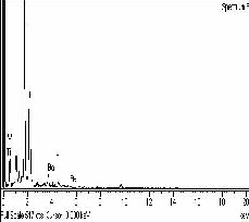
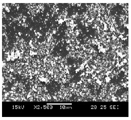
Scanning electron microscope is a promising technique for the topography study of thin film samples, as it provides valuable information regarding the size and shape of the particles or grains and also gives the information about the growth mechanism. Figure 2 shows scanning electron micrograph and EDAX image of as deposited BaTiO3 thin films. It can be seen that the fine grains with different sizes distributed the whole surface of the film. The EDAX peaks conforms the presence of barium, titanium and oxygen in the thin films. The maximum peak in the EDAX is due to the glass substrate used in the analysis.
The SEM image and EDAX spectra of CaTiO3 thin film deposited on glass substrate is shown in figure 3. This seemed to compose of domains formed by a large number of closely packed clusters covered on the substrate surface appears to be uniform. The EDAX patterns conforms the calcium, titanium and oxygen elements present in the coated substrates.
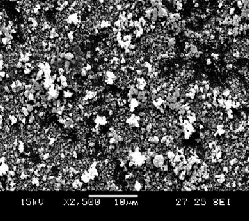
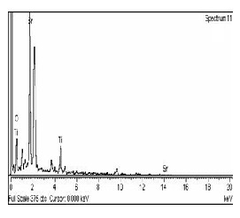
Figure 4 represents the SEM studied and EDAX of SrTiO3 thin film. The SEM study shows the formation of uniformly distributed nanocrystalline grains over the entire surface of the substrate. The EDAX spectrum shows the strontium,
titanium and oxygen elements were presence in the nanocrystalline thin film.
(a)

IJSER © 2014 http://www.ijser.org
(a)
International Journal of Scientific & Engineering Research, Volume 5, Issue 7, July-2014 591
ISSN 2229-5518
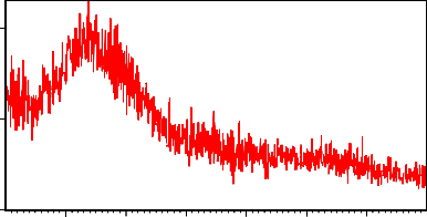
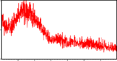
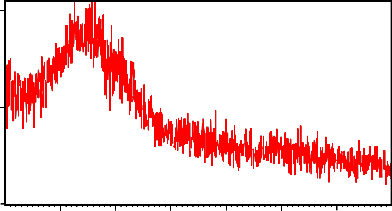
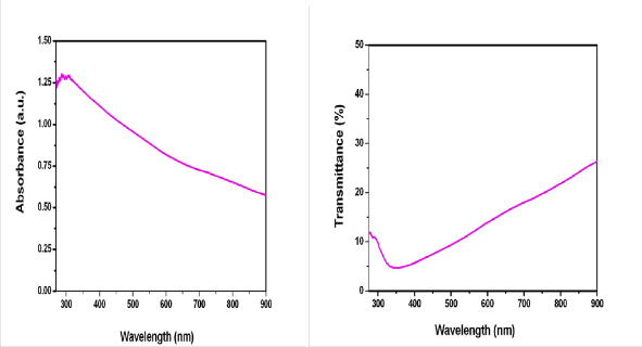
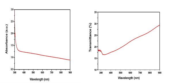
The figures 5 (a - b), 6 (a - b) and 7 (a - b) shows
the absorption and transmission spectrum of the barium titanate, calcium titanate and strontium titanate nanocrystalline films.
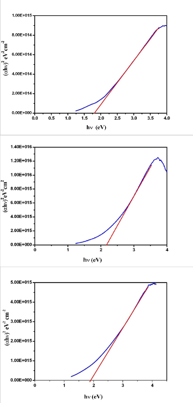
The optical absorption spectrum of these three titanates thin films on glass substrates were studied at room temperature in the wavelength range of 250 nm–800 nm. The absorption spectrum shows absorbance in the UV region and from the transmittance spectrum, the transmission of the film is 45% in the UV region. The optical absorption data of barium titanate, calcium titanate and strontium titanate were analyzed using the classical relation for near edge optical absorption of semiconductors [14 - 15]
αhν = A (hν – Eg)n
where ‘hν’ is the photon energy, ‘Eg’ is the band gap and ‘A’ is the constant. The ‘n’ is the equation has values 1/2, 2, 3/2 and
3 for allowed direct, allowed indirect, forbidden direct and forbidden indirect transitions [16 - 18] respectively.
The usual method for the determination of the values of (αhν)2 versus hν which was used to estimate the optical band
gap of barium titanate, calcium titanate and strontium titanate thin films. The estimated band gap values are 1.8 eV for BaTiO3,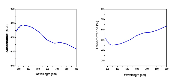
2.17 eV for CaTiO3 and 1.9 eV for SrTiO3, shows in the figure 8 (a - c).
IJSER © 2014 http://www.ijser.org
International Journal of Scientific & Engineering Research, Volume 5, Issue 7, July-2014 592
ISSN 2229-5518
(a) (b)
IJSER © 2014 http://www.ijser.org
International Journal of Scientific & Engineering Research, Volume 5, Issue 7, July-2014 593
ISSN 2229-5518
(a)
(b)
(c)
Barium, Calcium and Strontium titanate thin films were successfully prepared by chemical bath deposition technique. The
prepared BaTiO3, CaTiO3 and SrTiO3 thin films were thoroughly studied by various characterization techniques aimed at understanding the samples structural and optical properties. In the XRD pattern of samples, there is no signature of impurity
IJSER © 2014 http://www.ijser.org
International Journal of Scientific & Engineering Research, Volume 5, Issue 7, July-2014 594
ISSN 2229-5518
peaks. The EDAX confirmed the elements present in appropriate proportion. . From the UV study the band gap of Barium titanate, Calcium titanate and Strontium titanate were 1.8eV, 2.17eV and 1.9eV.
1. (a) W.H. Ma, C. Harnagea, D. Hesse, U.Gösele., Well-ordered arrays of pyramid-shaped ferroelectric BaTiO 3 nanostructures, Appl.
Phys. Lett., 83, (2003) 3770 - 3772.
(b) S. Mathews, R. Ramesh, T. Venkatesan, Benedetto., Ferroelectric Field Effect Transistor Based on Epitaxial
Perovskite Heterostructures, J. Science, 276, (1997) 238 - 240.
(c) B.H. Park, B.S. Kang, S.D. Bu, T.W. Noh, J. Lee, W. Jo., Lanthanum-substituted bismuth titanate for use in non- volatile memories Nature, 401, (1999) 682 - 684.
(d) M. Alexe, A. Gruverman, C. Harnagea, N.D. Zakharov, A. Pignolet, D. Hesse, J.F. Scott., Switching properties of
Self- Assembled ferroelectric memory cells, Appl. Phys. Lett., 75, (1999) 1158 - 1160.
(e) P.R. Evans, X.H. Zhu, P. Baxter, M. Mcmillen, J. Mcphillips, F.D. Morrison, J.F. Scott, R.J. Pollard, R.M. Bowman, J.M. Gregg., Toward Self-Assembled Ferroelectric Random Access Memories: Hard-Wired Switching Capacitor Arrays with Almost Tb/in.2 Densities, Nano Lett., 7, (2007) 1134 - 1137.
2. M. Cardona., Optical Properties and Band Structure of SrTiO 3 and BaTiO 3 , Phys. ReV. 140, (1965) A651 - A655.
3. K. Domen, A. Kudo, T. Onishi, N. Kosugi, H. Kurod., Photocatalytic decomposition of water into hydrogen and oxygen over nickel (II)
oxide-strontium titanate (SrTiO 3 ) powder. 1. Structure of the catalysts, J. Phys. Chem., 90, (1986) 292 - 295.
4. M.S. Wrighton, A.B. Ellis, P.T. Wolczanski, D.L. Morse, H.B. Abrahamson, D.S. Ginley., Strontium Titanate
Photoelectrodes. Efficient photoassisted electrolysis of water at zero applied potential, J. Am. Chem. Soc., 98, (1976)
2774 - 2779.
5. S. Burnside, J.E. Moser, K. Brooks, M. Gra¨tzel., Nanocrystalline Meaoporous Strontium Titanate as photoelectrode
Material for photosensitized solar devices: Increasing photovoltage through Flatband potential Engineering, J. Phys. Chem. B, 103, (1999)
9328 - 9332.
6. (a) H. Kato, A. Kudo., Visible-Light-Response and Photocatalytic Activities of TiO 2 and SrTiO 3 Photocatalysts
Codoped with Antimony and Chromium, J. Phys. Chem. B., 106, (2002) 5029 - 5034.
(b) R. Konta, T. Ishii, H. Kato, A. Kudo., Photocatalytic Activities of Noble Metal Ion-doped SrTiO 3 under Visible
Light Irradiation, J. Phys. Chem. B, 108, (2004) 8992 - 8995.
7. M. Miyauchi, M. Takashio, H. Tobimatsu., Photocatalytic activity of SrTiO 3 codoped with nitrogen and lanthanum under visible light illumination, Langmuir, 20, (2004) 232 - 236.
8. J.L. Giocondi, G.S. Rohrer., Spatial Separation of Photochemical Oxidation and Reduction Reactions on the Surface of Ferroelectric
Barium Titanate, J. Phys. Chem. B, 105, (2001) 8275 - 8277.
9. V.V. Lemanov, A.V. Sotnikov, E.P. Smirnova, M. Weihnacht., From incipient ferroelectricity in CaTiO 3 to real ferroelectricity in Ca 1−x Pb x TiO 3 solid solutions, Appl. Phys. Lett., 81, (2002) 886 - 888.
10. (a) P.K. Dutta, J.R. Gregg., Hydrothermal synthesis of tetragonal barium titanate (BaTiO 3 ), Chemistry of Materials, 4, (1992) 843 - 846.
(b) M.H. Um, H.J. Kumazawa., Hydrothermal Synthesis of Ferroelectric barium and strontium
titanate extremely fine particles, Mater. Sci., 35, (2000) 1295 - 1300.
(c) E.K. Nyutu, C.H. Chen, P.K. Dutta, S.L. Suib., Effect of microwave frequency on Hydrothermal Synthesis of
Nanocrystalline Tetragonal Barium Titanate, J. Phys.Chem. C., 112, (2008) 9659 – 9667.
11. G. Pfaff., Sol–gel synthesis of strontium titanate powders of various compositions, J. Mater Chem., 3, (1993) 721 - 724.
12. Y.F. Li, Q.Y. Lai., Study on Synthesis of Nano-crystalline SrTiO3 by Inverse Micell Microemulsion Method, Chin. J.
Inorg. Chem., 21, (2005) 915 - 918.
13. Y.B. Mao, S. Banerjee, S.S. Wong., Large-scale synthesis of single-crystalline perovskite nanostructures, J. Am.
Chem.Soc., 125, (2003) 15718 - 15719.
14. T. Dhannia, S. Jayalekshmi, M. C. SanthoshKumar, T. PrasadaRao and A. ChandraBose, Effect of Aluminium
Doping and Annealing on Structural and Optical Properties of Cerium Oxide Nanocrystals, Journal of Physics and
Chemistry of Solids, 70 (11), (2009) 1443 - 1447.
15. S. Varghese, M. Iype, E. J. Mathew and C. S. Menon, Determination of the Energy Band Gap of Thin Films of Cadmium Sulphide, Copper Phthalocyanine and Hybrid Cadmium Sulphide/Copper Phthalocyanine from Its Optical Studies, Materials Letters, 56 (6), (2002) 1078 - 1083.
16. A. F. Khan, M. mehmood, A. M. Rana and T. Muhammad, Effect of Annealing on Structural, Optical and Electrical
Properties of Nanostructured Ge Thin Films, Applied Surface Science, 256 (7), (2010) 2031 - 2037.
17. J.W. Jeon, D.W. Jeon, T. Sahoo, M. Kim, J.H. Baek, J. L. Hoffman, N. S. Kim and I.H. Lee, Effect of Annealing Temperature on Optical Band-Gap of Amorphous Indium Zinc Oxide Film, Journal of Alloys and Compounds, 509 (41), (2011) 10062 - 10065.
18. T. P. Kumar, S. Saravanakumar and K. Sankaranayanan, Effect of Annealing on the Surface and Band Gap
Alignment of CdZnS Thin Films, Applied Surface Science, 257 (6), (2011) 1923 - 1927.
IJSER © 2014 http://www.ijser.org