International Journal of Scientific & Engineering Research, Volume 2, Issue 3, March-2011 1
ISSN 2229-5518
Study of Microstructure and Mechanical
Properties of Human Cortical Bone
S. Biswas, P. C. Pramanik, P. Dasgupta, A. Chanda
—————————— • ——————————
one is a highly hierarchical structure and its mechani- cal properties are dependent on its structure and composition. Previous studies have focused on the macroscopic properties and their changes with respect to age and diseased condition. One such study showed with increasing tissue age, mineralization increases at certain portions and these hypermineralised portions become stiffer to some extent [1]. In another study about the post yield behavior it was found that middle-aged bone spe- cimens demonstrated higher yield strain and yield stress than those from the elderly ones [2]. Recent studies are focusing on the microstructural effect [3], [4]. Due to the presence of haversian canals as an integral part of its structure, bone is quite porous in nature. Studied have been done to find out the effect of porosity on mechanical properties [5]. A significant negative correlation was found between the elastic anisotropy and porosity of cor- tical bone [6]. Density also varies from person to person and also with age. Osteoporosis has become a serious issue all over the world and efforts have been made to predict the risk of fracture through different techniques [7], [8]. Nanoindentation studies have also become popu- lar for determining the hardness and elastic modulus of bone [9]. Variation of properties at specific sites like la- mella under different physiological conditions was also studied by few people [10]. Fracture toughness was another area of interest for many researchers who tried to correlate it with age and microdamage accumulation [11], [12]. In one of the very recent studies with the help of fi- nite element models and wavelet transforms, complete mapping of the energy descipation in bone after nanoin- dentation was shown. [13]. Laser engineered net shaping (LENS) is a rapid prototyping technique in which the ma- chine gets instructions from CT Scan data and can pro- duce or mimic a part of bone with powdered materials and without using any other tools. Very recent studies indicate that the in vivo life of implants depends on the porosity and mechanical properties of the material which should match with that of bone [14]. In this study we
have tried to map the apparent density and the porosities of different parts of femur both in males and in females. The load-deformation curve was also studied in details for both male and female under compression and tension to get the idea of its mechanical properties and the unde deformation of osteons under stress field was observed. Hardness was measured in both males and females throughout the femur shaft and its anisotropy was ob- served.
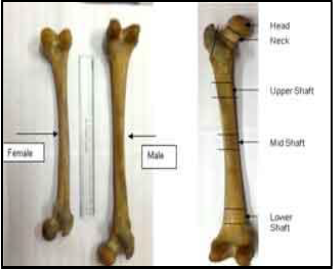
Femurs from both male and female human cadavers were collected, pretreated and cleaned. Density and porosity of the whole femur (shown in Fig-1a) were calculated by applying Archimedean principle. Then after drying they were sectioned into different parts as shown in Fig-1b.
Fig. 1. Human Femur and the different parts in which it was sectioned for measurements.
IJSER © 2011
2 International Journal of Scientific & Engineering Research, Volume 2, Issue 3, March-2011
ISSN 2229-5518
Then density and porosity of individual parts were al- so measured in the same way. A variation between the parts was very distinct. For observing the mechanical properties, samples of different sizes were cut and pre- pared following the ASTM standard (ASTM F 451
RE).The load-deformation curve of the cortical parts was obtained for both tension and compression from Univer- sal Testing Machine (UTM: INSTRON 4204, U.K.). Young’s modulus and work of fracture was calculated from the load-deformation curve itself. The loading was done till fracture for few samples, while in others it was stopped at various intermediate points, e.g. in the linear zone, in the plastic flow zone and then at the point of frac- ture to observe the deformation of the osteons at different stages of loading. This was mainly performed under compression as preparation of samples for tensile testing was little bit difficult due to some machine constraints. Experiments were also performed at different crosshead speeds of UTM i.e., with varying strain rate to determine its effect on stiffness of the bone. The cross section of the samples was polished in the polishing machine (LECO Spectrum System 1000, USA) with different grits until the value for the average centerline reached around 0.2534 micro meters.
Hardness was measured in both longitudinal and transverse direction to check the anisotropy using Vick- er’s Hardness testing machine (LV 700AT, LECO, USA). The structures of osteons were observed from the cross sectional images of the cortical bone (of both male and female) by a Reflecting inverted optical microscope (Olympus GX51, Japan). Using image analysis software (Analysis five, Olympus, Japan) the aspect ratio, lamellar thickness, diameter of haversian canals were calculated.
The structure of bone was thoroughly observed under the optical microscope under different magnifications. Os- teons were clearly visible in the polished cross sections of the cortical part. Osteonal density was slightly different in different regions of the cross section. The average number of osteons per mm2 was nearly 19 (1 .9 x 10-5 per sq. mi- cron) in the inner part (towards the bone cavity) to about
14 (1.3 x 10⁻⁵ per sq. micron) in the outer part (Fig-2). With increasing magnification, the lamella, which are the con- centric layers of tissue around the Haversian canal, were clearly visible. The thickness of the lamella ranged from
6.25 micron to 10.55 micron from inward to outward with a scatter of ±10%. Almost all the osteons had a combina- tion of thin and thick lamella. The diameter of the Have- sian canal had an average value of 78.18 micron towards the periphery as compared to 54.66 micron in the inner part with a large scatter in the values all over the region. Towards the inner side, the osteons were found to be more closely spaced which may be indicative of compara- tively higher rate of bone formation in that region. Pic- tures taken from the scanning electron microscope re- vealed even minute structures called the lacune which are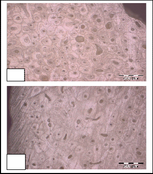
the sites for the bone cells (osteocytes).
a
b
Fig. 2. Cross section of cortical bone (a) inner part (b) outer part man
The lacunae were visible along the layers of lamella. In- terconnections between them are established by the cana- liculi. Thus the structure reveals that such discontinuities are integral part of the bone structure. It will be shown later how they contribute to the fracture of bone by pro- moting the coalescence of cracks under different types of loading. Therefore concentration of porosity is to be taken into consideration when properties of bone are con- cerned. We measured the porosity of the whole femur and found it different when compared to the porosity of different parts of the same femur considered separately. Fig-3 shows the porosity distribution of in two male and two female samples. The cortical part is quite less porous than the head or the trabecular part in general. The densi- ty in females was found to be slightly lower in males as shown in Fig-4.
Fig. 3. Porosity distribution in Femur
IJSER © 2011
International Journal of Scientific & Engineering Research, Volume 2, Issue 3, March-2011 3
ISSN 2229-5518
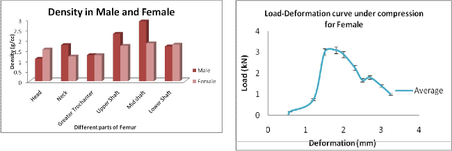
Fig. 4. Density distribution in male and female femur
But the results of a students t-test performed on both the sets of data showed that the difference in males and females were not significant at 95% confidence level. In our samples, porosity of one female femur was extremely high than others. This may be due to increased suscepti- bility to osteoporosis at higher age.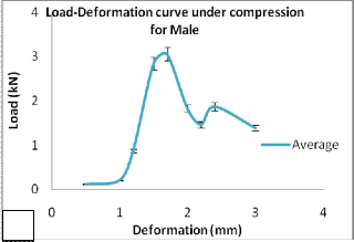
The load-deformation curves were obtained from compressive and tensile loading to study the mechanical properties. The initial part showed a stunning similarity for all the samples both in male and female. The load- deformation curve could be divided into two zones i.e., the elastic zone which was linear in all cases, and a nonli- near zone. Under compression, after reaching the peak load the curve was more flat in females than in males. The nonlinear zone revealed number of kinks representing initiation of fracture, progressive fracture and finally complete breakage occurred with a comparatively sharp drop of load. However during experiment, at these in- termediate points no major splitting was observed, there were some primary signs of cracking. The parts of load- deformation curve corresponding to fracture zone were not truly identical in all the cases but the general pattern was comparable. Figure-5 shows a representative curve for the load deformation curves in both male and female with error bars showing 5-10% scatter at each data point.
a
Fig. 5. Representative Load-Deformation curves a) For Male samples b) For Female samples of Femur
The equations fitting the individual curves of these two zones are given in the Table1. In the elastic zone, they are all fitted into linear equations with R2>0.95 indicating a good amount of fitting, although slopes may have been different. For the non-linear part, most of the curves fitted with a 2nd degree polynomial and few with 3rd degree, all having R2 values greater than 0.9. The ultimate strength was in the order of 158.66 MPa with standard deviation of
5.9 in females and 150.76 MPa with standard deviation of
11.9 in males. The elastic modulus was also calculated.
The stiffness value determined in our study ranged from
10 to 15 GPa which was also verified by the nanoindenta- tion technique. Work of fracture, which is the energy ab- sorbed by the bone before complete fracture was obtained separately from the area under each curve and was
slightly more in females in most of the cases than in males.
TABLE 1
EQUATIONS DESCRIBING THE LINEAR AND NON-LINEAR PART
OF THE LOAD-DEFORMATION CURVES
IJSER © 2011
4 International Journal of Scientific & Engineering Research, Volume 2, Issue 3, March-2011
ISSN 2229-5518
The difference in the average values of work of frac- ture has been around 10% which is well within possible experimental scatter. Rather we should say the values were quite comparable. This strongly contradicts the pre- vailing hypothesis of higher vulnerability of female bone due to increased porosity particularly at higher age. The samples taken were rectangular in shape and in case of both tensile (Fig-6a) and compressive (Fig-6b) fracture it was observed that the fractured surface made an inclina- tion with the vertical axis. The crack propagated making almost a 30o angle with the vertical axis of the bone.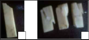
b
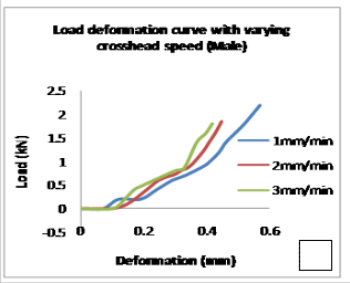
Fig. 7. Load-Deformation curve with varying cross head speed
(a) for Female (b) for Male
a b
Fig. 6. Fracture plane making an angle with the vertical axis
(a) under tension (b) under compression
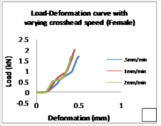
In few cases of compressive loading for female bone samples, the samples experienced a crushing pattern where instead of forming asharp edge it got smashed from the top up to the bottom surface. The slope of the load-deformation curve changed as the crosshead speed was varied from 0.5mm/min to 4 mm/min. It was thus in accordance with the previous findings that stiffness of the bone increases with increasing loading rate or strain rate in case of tensile loading [15].Fig-7 shows the variation of stiffness with varying crosshead speed, or in other words varying strain rate. Thus from this figure it can be ob- served that not only under tension,
a
effect of varying strain rate on the slope of the load- deformation curve was prominent in case of compressive loading also.
SEM images (Fig-8) of the fractured surface both under compression and tension showed the deformation of os- teons very clearly. In case of compressive fracture, the osteons deformed to such an extent that the aspect ratio of the osteons reached about 1:2. But in case of tensile frac- ture, osteons were seen to be protruding from the surface like tubular structures without much change in the aspect ratio. Again, fractography showed the fracture in the ce- menting zone appeared to be comparatively smooth or cleavage in nature in contrast with fibrous fracture in la- mellar zones. As observed in case of porosities, for other mechanical properties also, there is a high chance that their values will differ when the whole bone is considered than when different parts are considered separately.
Hardness in the cortical part along the femur length did not vary significantly. But values of the indentations over the lamella were little higher (around 0.38 GPa) than those in the cementing zone (0.33GPa). The hardness of the cortical part of female bones was found to be lower as compared to males. Not much difference could be ob- served in longitudinal and transverse values. More or less it was observed that the microstructural features like la- mella and interstitial zone had some effect on the hard- ness and also on crack propagation. Also the typicality in fracture and deformation of osteons under various types of loading had been identified quite clearly but the quan- titative estimate of their (collagenous lamella or minera- lised interstitial zone) individual contribution in control- ling load-deformation as well as minute fracture events is yet to be understood in a comprehensive manner. It needs controlled set of experimentation, that are being done at present.
IJSER © 2011
International Journal of Scientific & Engineering Research, Volume 2, Issue 3, March-2011 5
ISSN 2229-5518
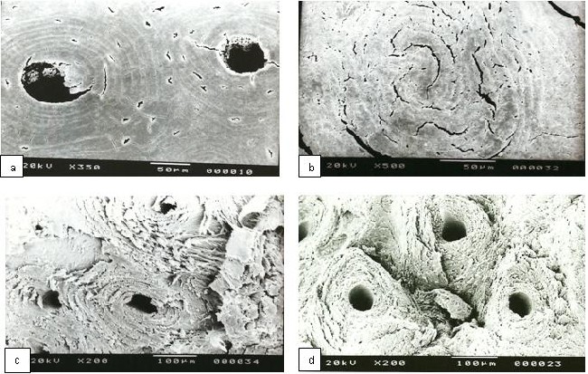
Fig. 6. SEM images of (a) undeformed osteons (b) Cracks developed along the lamellae well as across it.(c) Surface under compressive fracture (d) Surface under tensile fracture
From the study done so far, it has been observed specifi- cally that the osteons suffer different types of damage and fracture in tensile and compressive loading. More defor- mation of microstructure was found in compression while in tension, there were more signs of shearing and tearing in-between the osteons. From the load-deformation curves it was evident that both male and female bones behaved similarly before yield point but post yield cha- racteristics were different. It may be attributed to more energy absorption capacity of female bones. Although the lamellar structure is known to provide ductility to bone, cracks propagating across the lamella under high stress may be indicative of some inherent brittleness of bone. Finally it may be noted that mechanical behavior of only the mid-shaft cortical part is considered here for detailed study, the other portions are also to be studied before we make any generalization about the micro-structure- property relationship of bone.
The authors acknowledge sincere support and assistance from all the technical staff of the School of Bioscience and Engineering, Jadavpur University. We express our grati- tude to Sri Ramprasad Das for his technical help in cut- ting the bones.
[1] J.Y. Rhoa, P. Ziouposb, J.D. Curreyc, G.M. Pharr. Microstructural elas- ticity and regional heterogeneity in human femoral bone of various ages examined by nano-indentation. Journal of Biomechanics 35 (2002)
189–198.
[2] Huijie Leng, X.Neil Dong, XiaoduWang. Progressive post-yield beha- vior of human cortical bone in compression for middle-aged and elder- ly groups. Journal of Biomechanics 42 (2009) 491–497
[3] Jeffry S. Nyman, Michael Reye, Xiaodu Wang. Effect of ultrastructural changes on the toughness of bone. Micron 36 (2005) 566–582
[4] R.K. Nalla a, J.J. Kruzic b, J.H. Kinney c, M. Balooch c, J.W. Ager III a, R.O. Ritchie,a. Role of microstructure in the aging-related deterioration of the toughness of human cortical bone. Materials Science and Engi- neering C 26 (2006) 1251 – 1260
[5] Igor Sevostianov, Mark Kachanov. Impact of the porous microstructure on the overall elastic properties of the osteonal cortical bone. Journal of Biomechanics 33 (2000) 881-888
[6] X. Neil Dong1, X. Edward Guo. The dependence of transversely iso- tropic elasticity of human femoral cortical bone on porosity. Journal of Biomechanics 37 (2004) 1281–1287
[7] M. Muller, D. Mitton, P. Moilanen, V. Bousson, M. Talmant, P. Laugier.
Prediction of bone mechanical properties using QUS and pQCT: Study
of the human distal radius.
[8] G. Dougherty. Quantitative CT in the measurement of bone quantity and bone quality for assessing osteoporosis. Medical Engg Physics Vol.18 no. 7 pg 557-568. 1996.
[9] Philippe K. Zysset, X. Edward Guo, C. Edward Hoffler, Kristin E.
Moore, Steven A. Goldstein. Elastic modulus and hardness of cortical
and trabecular bone lamellae measured by nanoindentation in the hu- man femur. Journal of Biomechanics 32 (1999) 1005-1012
[10] S. Hengsberger, A. Kulik, and PH. Zysset. Nanoindentation Discrimi- nates the Elastic Properties of Individual Human Bone Lamellae Under
IJSER © 2011
6 International Journal of Scientific & Engineering Research, Volume 2, Issue 3, March-2011
ISSN 2229-5518
Dry and Physiological Conditions. Bone Vol. 30, No. 1.January
2002:178–184
[11] Y. N. Yeni and T. L. Norman. Fracture Toughness of Human Femoral Neck: Effect of Microstructure, Composition, and Age. Bone Vol. 26, No. 5 May 2000:499–504
[12] R.K.Nalla, J.J.Kruzic, J.H.Kinney, R.O.Ritchie. Effect of aging on the toughness of human cortical bone: evaluation by R-curves. Bone 35 (2004) 1240 – 1246
[13] Kuangshin Tai1, Ming Dao1, Subra Suresh, Ahmet Palazoglu And Christine Ortiz. Nanoscale heterogeneity promotes energy dissipation in bone. Nature Materials VOL 6 JUNE 2007
[14] Peter Zioupos a, Ulrich Hansen b, John D. Currey. 2008, Microcracking damage and the fracture process in relation to strain rate in human cor- tical bone tensile failure. Journal of Biomechanics, 41, 2932–2939
IJSER © 2011