In this project, the test results indicated that the setting
(block size= 2x2, window size= 18x18 & multiplication fac-
es.
The opening morphological operation gives more acceptable
tor α = 0.025) led to best thresholding results.

International Journal of Scientific & Engineering Research, Volume 5, Issue 4, April-2014 1026
ISSN 2229-5518
Palm Vein Recognition and Verification System
Using Local Average of Vein Direction
Asmaa M.J. Abbas and Dr. Loay.E. George
Abstract— During the last years, hand vein patterns recognition is one of the most recent biometric technologies used for the identification/verification of individuals. The vein trace is hard to damaged, changed or falsified since veins are internal to the human body. In this paper a novel palm vein recognition and verification system is presented. The first step in the proposed system is image enhancement and localization of veins grid which is a major challenge due to poor quality of veins images. The second challenging task is the palm vein feature extraction. In this research the spatial distribution of local averages of veins direction is introduced and used as feature vector.
The system was tested over a database collected from 250 volunteers, where 24 images for the 2 palms are collected for each person. In total, a database contains 6,000 images belong to 500 different palms. The attained identification result is encouraging (99.95%). The verification tests indicated the achieved minimum equal error rate (EER) is 0.24%.
Index Terms— palm veins, Identification, Verification, Recognition, Biomatric, Pattern Recognition.
—————————— ——————————
he prime responsibility of any technological development, con- cerned with access control issue, is to provide a unique and se-
cure identity for citizens, customers or stake holders, and it is a ma- jor challenge for organizations. There is an increasing interest for biometric in the research community since the traditional verification methods (such as passwords, personal identification numbers (PINS), magnetic swipe cards, keys and smart cards) offer limited security and are unreliable [1].
Biometrics means "life measurement". A biometric system ei- ther makes identification or verifies an identity. It is based on the use of unique and measurable physiological or behavioral characteristics. Physiological characteristics include, but are not limited to, a per- son’s vein patterns, facial structures, ocular characteristics, hand geometry, or fingerprint [2].
Each kind of physical biometrics has merits and demerits. In the case of fingerprints, direct contact of the finger with the finger- print-image-extracting sensor causes degradation in performance, where good-quality fingerprints are hard to obtain due to oil from the finger, moisture, dirt,...etc. For retina scanning users must place their eye close to the scanner, causing an uncomfortable feeling and con- cerns of privacy. With hand-shape recognition devices, problems may arise with users who suffer from arthritis or rheumatism, leading to poor performance.
scbaghdad.edu.iqCompared with the other physical characteristics, vein pattern recognition is one of the newest biometric techniques were developed to resolve many problems facing the traditional bi-
———————————————
• Dr. Loay.E. George is currently computer deparment chief in college of
Science in Baghdad University, Iraq. E-mail: Loayedwar57@yahoo.com
ometric systems. The main reasons for adopting vein palm biometric techniques are:
1. The acquisition process of palm vein image needs no direct con- tact with the vein pattern-extracting sensor. Since contactless models are more hygienic than all forms of contact biometrics, so the user comfort is improved with use of vein imaging technolo- gy [3].
2. Vein pattern does not change over time [4], and they can repre- sent the liveness of a person [5], so the cognition performance can be improved with use of vein imaging technology, and a sta- ble operation is expected [3].
3. Vein recognition technology is notably less costly than many of other biometric technologies (like, iris scanning technology) [3]. In fact, the only biometric solution less expensive than palm-vein is fingerprint recognition but it has its own overheads on security feature [1].
4. For the case of veins imaging, in addition the blood vessels are hidden underneath the skin and are mostly invisible to the human eye; the vein patterns are much harder for intruders to copy, and extremely difficult to steal/misuse compared to other biometric features [6].
Hand veins biometric are robust and steady human authentica- tion more than other biometric technologies so it is considered to be one of the most reliable biometrics for personal identification [4].
Many recognition and verification technologies using bi- ometric features of hand veins have been developed over the few last years.
Heenaye and Ali [6] introduced a veins recognition method based on quadratic inference function to extract the
IJSER © 2014 http://www.ijser.org
International Journal of Scientific & Engineering Research, Volume 5, Issue 4, April-2014 1027
ISSN 2229-5518
dorsal hand vein features. For matching task they used the
Euclidean distance measure.
Wang and et al [7] presented a new method based on Par- tition Local Binary Pattern (PLBP). After preprocessing, the image is divided into sub-images. A set of LBP uniform pat- tern features is extracted from each sub-image. Then, these sets are combined to form the feature vector for token vein texture features. Wang and etal [8]proposed a novel approach to extract multi-scale LBP features of hand vein images using wavelet decomposition. Wang and Chen [4]setup a creatively vein-image capturing system and presented a novel frame- work. It is composed of image enhancement, feature extrac- tion, noise removal, thinning, skeletonization, and pruning for vein pattern extraction.
Liu and Zhang [9] presented a new method palm recogni- tion based on Two-Dimensional FLD (2DFLD). They applied PCA, PCA+FLD and 2DFLD algorithms to extract the palm- dorsa vein feature subspace.
Wang and et al [10] introduced a novel method for hand vein recognition based on fusing multiple sets of key points extracted from the scale-invariant feature transform (SIFT).
Tang and et al [11] proposed a novel approach for hand dorsa vein recognition; it makes use of multi-level key point detection and SIFT feature based local matching. Prabu and Sivanandam [12] attempted to improve the performance of palm vein based verification system with the help of energy
feature based on wavelet transform.
The layout of the proposed system is shown in Figure(1). It is consist of three main modules: preprocessing, feature extraction and matching. Detail descriptions of the system modules are given in the following sections.
Fig.(1) The general structure of the proposed system
2. Brightness Stretching & Normalization: A simple linear type of contrast stretching is applied to enhance the visual appearance of the image details. The dynamic range of pixels values is adjusted to be [0,1]. This process is done using the following equation [13]:
N − N
The performance of feature extraction algorithm relies heavily on the quality of the input images. In practice, the quality of the ob- tained raw images is very low because the images are blurred and noisy due to variations in environmental conditions, skin conditions, and acquisition devices, etc. A set of tasks are applied; they are nec- essary to improve the clarity of the vein pattern structure and localize the veins grid.
A. Image Enhancement
The purpose of this stage is to improve the imaging quality so that vein patterns can be more easily detected during the segmenta- tion. This stage implies the following steps:
1. Image Preparation: The input image is converted to be8-bit gray
image. Then, it is converted to the negative which make the ROI
as bright region.
N ( x, y) = max min (O( x, y) − Omin ) + Nmin
Omax − Omin
3. De-Noising & Integration: Despite the image is blurred, a simple mean smoothing filter is used to reduce the noise and to integrate the white ROI. Mean filter can lead to good result, when apply- ing it in a specific way. The size of the applied mean filter is 7x7, and is applied four times to obtain an acceptable result, denoted Is ().
B. Segmentation
Once the noise is reduced and the contrast enhanced, segmenta- tion permits to separate the vein pattern from the background, it con- sists of the following tasks:
1. Thresholding: Because the images contain considerable back- ground noise, and there is variation in contrast and illumination gradient. So, the local thresholding mechanism is more suitable to be used than the global thresholding. In this project, the ap- plied local thresholding process implies the following steps:
• Partition the image, Is (),into small non-overlapped blocks,
each has size (kxk).
IJSER © 2014 http://www.ijser.org
International Journal of Scientific & Engineering Research, Volume 5, Issue 4, April-2014 1028
ISSN 2229-5518
• For each block, make a scanning window (with area nxn) co- vers the block area and extends to the surrounding area (i.e., n>k), see figure (2).
• Determine the mean (m) and standard deviation (σ) of the
pixels values located inside the window (nxn).
• Then for each pixel belong to the scanned block (mxm) apply the following thresholding criterion:
smoothing the contour of the objects (veins), (4) solving the problem of existence of regions containing mixed white and black pixels, (5) removing small holes, and (6) conserving the necks and slim parts of veins from breakage. When applying closing morphological opera- tion on the veins images several defects may appear in the resulting images, like: (1) the small objects are kept and strengthened instead of removing them from the image, (2) when two veins are too close to each other they may linked, (3) misclassification may occur for
Is (x, y)
Ithr (x, y) =
if Is (x, y) ≥ m − ασ
otherwise
the regions containing mixed white and black pixels, because such regions are always considered as veins. Because of the above rea- sons, the closing operation is not useful to improve the studied imag-
In this project, the test results indicated that the setting
(block size= 2x2, window size= 18x18 & multiplication fac-
es.
The opening morphological operation gives more acceptable
tor α = 0.025) led to best thresholding results.
Fig.(2) The Scanning Block and W indow Areas
results than the closing operation for allocating the shape of veins in
the image. It is used to remove small objects from the image without altering the overall shape and size of the large objects (veins object). Also, its smooth the contours of the existing large objects. But when the traditional opening operation is applied on the veins images sev- eral defects appeared, like: (1) the vein in some places appears too skinny, or it may be narrow (like a neck) and due to traditional open- ing operation this part will disappear, (2) there are many regions in the vein image are mixture of white and black pixels; these regions may be parts of veins, parts of the background, or noise; they appear due to converting the image from gray to the binary. The traditional opening operation deletes all these regions without any consideration to the probability of being veins, (3) the holes are not removed from the veins regions. These defects significantly affect shape of veins grid.
2. Binarization: After applying local thresholding the produced vein image will have better background brightness, and in such case global thresholding becomes more suitable to do binariza- tion. In our proposed system the following steps are adopted to do image binarization:
• Get the highest pixel value (Max) found in the threshold im-
age.
• Map each pixel belong to threshold image using the follow- ing:![]()
I ' ( x, y) = I ( x, y) × 255
thr Max
• Binarize the pixels values using the following criterion
For these reasons, a new algorithm is proposed to allocate and improve the shape of veins grid by cleaning the appeared gaps and pores from the image. This algorithm works to:
• Remove the small objects from the image.
• Smooth the boundary of veins (contour).
• Remove the small holes.
• Conserve the necks and slim veins regions from breakage defect.
• Take into consideration that the mixed regions could be part of the veins or not.
In the proposed algorithm the treatment of each pixel depends on its location. The applied steps of the introduced algorithm are:
1. For each corner pixel the algorithm counts the number of all the
adjacent white pixels; then the pixel is treated depending on this number. There are two possible cases: For black pixel (i.e., 0): if
0
I bin ( x, y) =
1
if I ' ( x, y) < T
otherwise
the number is more than one, then the pixel is considered as gap and converted to the white pixel, otherwise it kept black. For
The test results indicated that the best value for T is 100.
This stage was used to process the image after the segmentation, in order to reduce the effect of undesired elements such as noise, and improve the shape of the vein grid. It consists of the following steps:
I. Cleaning the Gaps and Pores: In this stage we need to reconstruct the vein image in order to improve the shape of the veins; this was accomplished by: (1) eliminating the small objects in the image; these objects are noise and parts of the background, they were classi- fied as veins due to misclassification, (2) eliminating protrusions, (3)
white pixel (i.e., 1): if the number is zero then pixel is consid- ered as pore point and converted to black, otherwise it is kept white.
2. For each pixel on the edge line the algorithm counts the number
of all adjacent white pixels, then, the pixel is treated depending on this number. The number of white pixels. There are two cas- es: For black pixel (=0): if the number is higher than two, then the pixel is considered as gap point and converted to white, oth- erwise it is left black. For white pixel (=1): if the number is less than two, then the pixel is considered as pore point and convert- ed to black, otherwise is left white.
IJSER © 2014 http://www.ijser.org
International Journal of Scientific & Engineering Research, Volume 5, Issue 4, April-2014 1029
ISSN 2229-5518
3. For each pixel in the inner region: the algorithm counts the number of all the adjacent white pixels then the pixel is treated depending on this count value. The result depend on the value of the selected pixel, there are two cases: For black pixel (0): if the
![]()
β > M 2 > α
M1
Where,
number is more than four, then the pixel is considered as gap point and converted to white, otherwise it is left black. For
M1 = ∑ ∑ I ( x, y),
∀y ∀x
M 2 = ∑ ∑ I ' ( x, y)
∀y ∀x
white pixel (1): if the number is less than four, the pixel is con- sidered as pore point and converted to black, otherwise is left white.
II. Thining: The vein patterns could have different thicknesses due to physiological status of a person (for example, fatigue or non-fatigue) or it may due to the preprocessing operations. Therefore, vein thick- ness is not a stable pattern for recognition. So, the thinning operation is needed to ensure representation of veins objects that can correctly describe the main features like shape and connectivity.
A new thinning method is developed for more control to make thinning up to a certain width. The proposed thinning algorithm is designed to reduce the width of the pattern to five pixels width line. It will solve the problem of hand shift when taking its IR image. The algorithm tests only the white pixels in the image, either it decides to
Where, I() is the input image array, I’() is the image array
after processing. In this project, the parameters values
(α=0.985 and β=1.015)led to the best skeleton result.
III. Fine Thining: The purpose of this stage is making better thinning for the veins body. More thinning can be achieved by applying the following steps:
1. Integration: In this stage, the integration process is applied
to make veins thinner; this is done by increasing the bright- ness of vein’s center, and reducing the brightness of the vein’s sides. This step will give more characterization for the center of the veins.
The integration process works by opening a window (nxn) around each pixel and calculate the new value by ap- plying the following equation:
leave the pixel or convert it to black pixel (white pixels indicate vein r r
and black pixels refer to background). The introduced algorithm consists of the following steps:
I ' ( x, y) = ∑ ∑ I ( x + i, y + j)
j = − ri = − r
1. Apply the mask, M(),on the selected pixel in order to find if the pixel is inside the vein area or not, this implies counting![]()
r = 1
2
(n − 1)
the number of the white pixels surrounding the tested white pixel; this is done by applying the following equation:
Where, I'() is the resulted integrated image, I() is the in- put image and n is the window length.
2 2 2. Edge Normalization: now, the normaliza-tion process is ap-
Fout ( x, y) = ∑ ∑ I ( x + i, y + j) M (i + 2, j + 2)
j = −2i = −2
Where, I() is the input image array, M() is the used mask (in our system a 5x5 mask is used because the required vein width is five pixels), Fout is the number of the adjacent white pixels. All the mask's element values are "1", except the cor- ner pixels they set "0". This mask is designed to make the
plied to improve the intensity contrast between the center of veins and its sides. In this project, the applied normalization process consists of the following steps:
• For each non zero pixel, open a window (nxn) to determine
the mean value (m) for all the pixels located inside the win- dow. Then, the following threshold-ing criterion is applied:
output vein as smooth as the natural veins. If the value of Fout
is more than 20 then the tested white pixel is considered as
I ( x, y)
I th ( x, y) =
0
if I ( x, y) ≥ αm
otherwise
part of vein and left as it is; otherwise the algorithm applies the multi-directional checks on that pixel.
2. In this step, the thinning operation starts after applying the full area test and knowing that the specific pixel is not inside the vein. A set of multi-directional checks are applied on the test-
In our applied system, when the window size is taken (3x3) and the multiplication factor α is set 0.25; it was found that the attained normalization is the best.
• Determine the new value using the following equation:
1
ed white pixel to decide if the pixel has to be removed (i.e.,
convert the white pixel to black) in order to thin the vein seg-
I R ( x, y)
![]()
= I th ( x, y)
m
ment. The multi-directional checking operations imply check- ing along the horizontal extent: for each tested pixel the algo- rithm scans the four neighbor pixels on both sides horizontally to check the existence of the four successive white pixels. Now, if the algorithm found these successive white pixels on
Then find the global maximum pixel value (I max ).
• Calculate the normalization value for each pixel by applying
the following equations:
I norm ( x, y) = Slp × I R ( x, y)
I m′ ax
one side or both sides it will start the vertical checking. Oth- erwise, the pixel is left as white pixel and considered as a part
Slp =
I
max × α
of vein skeleton. This task is repeated for other directions (i.e., vertical, main diagonal, and second diagonal).
3. Apply the gaps and pores algorithm twice.
4. The algorithm repeats its steps until the required thinning achieved. The proposed algorithm rounds are stopped when the following conditions satisfied:
IJSER © 2014 http://www.ijser.org
Where, IR () is the final process image array, Inorm () is the
edge normalized image array, I'max is set 255, Imax is the maximum found pixel value, and (α=0.9).
3. Binarization with Thinning: This stage aims to allocate the center of vein. In this stage, the gray image is converted to binary image to keep only the object of interest. Several
International Journal of Scientific & Engineering Research, Volume 5, Issue 4, April-2014 1030
ISSN 2229-5518
global and local adaptive thresholding methods were investi-
gated and founded that these methods lead to lose some parts of the veins.
In the proposed method each non-zero pixel in the image is compared twice with its four adjacent pixels. In the first check, the pixel is compared with the left and right pixels; and in the second check, it is compared with the up and down pixels, then by applying the following criteria the pix- el is binarized:
1 if
(I ( x − 1, y) ≤ I ( x, y) ≥ I ( x + 1, y))
Ibin ( x, y) =
0
or (I ( x, y − 1) ≤ I ( x, y) ≥ I ( x, y + 1))
otherwise
Where I()is the input gray image after normalization and Ibin ()is the output binary image. Figure (3) presents an illus- tration for the preprocessing stage.
In this stage, a set of key information is extracted from the final processed vein’s image. The extracted information represents the set of required features to distinguish between persons. The local aver- age of veins directions method is proposed as discriminating veins grid features.
The benefits of this method are: (1) it is applicable in spatial do- main and reflects the directionality of veins tracks, and (2) it is indi- rect measure to the distribution of veins density in each parts of im- age. So, this set of features depends mainly on the distribution of vein’s directions at different parts of the image.
The following steps have been applied to extract the features vec-
tor:
1. Determine the direction of each vein pixel in the image; that is by checking if the pixel is located in the vein body along the hori- zontal, vertical, main diagonal or second diagonal direction. This is done by checking all the connected pixels, surrounding the tested pixel, along the four directions and to count the extent of these pixels. Then, the longest extent is taken as the local direc- tion of the vein at the tested pixel location.
2. The resulting 2D-array of vein direction is divided into blocks.
3. The average of local directions is determined for each block, separately; and the determined average values for all blocks are assembled as a feature vector. Four features extracted from each block, that is the densities of the (i) vertical, (ii) horizontal, (iii) main diagonal, and (iv) second diagonal veins direction. Each of these direction features is calculated by counting the number of pixels which have that direction, and then dividing it by the total number of veins pixels. Figure (4) presents an example of pixel with second diagonal feature.
•
Fig (3) Illustration for the outcomes of preprocessing stage
IJSER © 2014 http://www.ijser.org
International Journal of Scientific & Engineering Research, Volume 5, Issue 4, April-2014 1031
ISSN 2229-5518
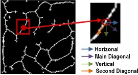
Fig. (4) Example of pixel with second diagonal feature
This stage calculates the degree of matching between two vein patterns. The extracted vein patterns of the input image can directly be compared with the stored templates. A similarity measure should be used to evaluate the similarity degree between a template and an input pattern.
In this work the two Euclidean similarity measures (i.e., mean square difference and the mean absolute difference; called city block distance) have tested to evaluate their suitability for matching the veins feature vectors.
The performance of the proposed system was tested using a da- tabase collected from 250 volunteers, including 195 males and 55 females. The age distribution of volunteers is from 17 to 60 years. The samples are collected in two separate sessions. In each session, the subject was asked to provide 6 images for each palm. Therefore,
24 images of each volunteer are collected, 12 of them for each of his/her 2 palms. In total, the database consists of 6,000 images taken from 500 different palms. The average time interval between the first and the second sessions was about 9 days. The proposed method have used the near-infrared (NIR) illuminations images of PolyU multi-spectral palm print database[14].
The results of the conducted tests are described in details in the following subsections.
To achieve an efficient performance for vein recognition and verification, veins grid must be extracted correctly. The localization of vein can be subjectively evaluated by matching the extracted vein grid with the veins network that can be seen in the original image. Figure (5) presents the final vein localization image for one person and his image after projecting the vein localization on the enhanced original images (note: the original images have been modified for the purposes of assisting the subjective comparison task).
In the identification mode, the system performance is measured us-
ing the parameter correct recognition rate (CRR); it is the ratio of the number of samples being correctly classified to the total number of tested samples.
Fig.(5) Final vein localization image for one person
There is a set of system parameters that affect the recognition performance behavior, the main affecting ones are :
• Number of blocks (ROI divided into NxN block).
• The ratio of the overlapped blocks.
The recognition rate is determined for the two cases: (i) apply thinning only, and (ii) apply fine thinning. The Euclidean distance and City Block distance are used to measure the similarity between features vector. Table (1) lists the attained recognition values for different number of blocks.
TABLE 1
RECOGNITION RATE VERSUS THE NUMBER OF BLOCKS
No. of Blocks | Recognition Rate | |||
No. of Blocks | With Thinning | With Fine Thinning | ||
No. of Blocks | Seq. Distance | Abs. Distance | Seq. Distance | Abs. Distance |
4x4 | 85.65% | 85.65% | 95.25% | 95.90% |
5x5 | 95.35% | 95% | 98.15% | 98.65% |
6x6 | 98.55% | 98.35% | 99.30% | 99.60% |
7x7 | 99.35% | 99.30% | 99.60% | 99.80% |
8x8 | 99.75% | 99.70% | 99.80% | 99.80% |
9x9 | 99.75% | 99.80% | 99.90% | 99.95 |
10x10 | 99.65% | 99.55% | 99.75% | 99.85% |
The table shows that the recognition rate is improved when adding the fine thinning stage. Also, according to the blocks number parameter, the best recognition rate is achieved when dividing the image into 9x9 blocks, while the second best result came when di- viding the image into 8x8 Blocks.
The performance of verification system is evaluated by the Re- ceiver Operating Characteristic (ROC) curve, which illustrates the False Rejection Rate (FRR) against the False Acceptance Rate (FAR) at different thresholds on the matching score[15]. The performance is also evaluated by the Equal Error Rate (EER), which is defined as
IJSER © 2014 http://www.ijser.org
International Journal of Scientific & Engineering Research, Volume 5, Issue 4, April-2014 1032
ISSN 2229-5518
the error rate where the FAR and the FRR are equal. The EER indi- cate the minimum verification error. So, the threshold value is select- ed according to the minimum error.
The FAR and FRR are defined, respectively, as[16]:
FRR = ![]() FAR =
FAR = ![]()
Also the performance of biometric systems can be measured by accuracy; i.e., the proportion of correct predictions, without consid- ering what is positive (P) and what is negative (N) [17].
Accuracy=(TP+TN) / (P+N)
Table (2) shows FAR, FRR and Accuracy values with different threshold.
TABLE 2
FAR,FRR AND ACCURACY VERSUS DIFFERENT THRESHOLD
TABLE (3) EXECUTION TIME
Stage | Time (msec) |
Preprocessing | 1.704 |
Feature Extraction | 170.14 |
Matching | 158.415 |
Total | 330.25 |
Many vein recognition methods have developed and published. Here, we give a comparison between the performance of our pro- posed method and some the published methods.
Table (4) presents the recognition(CRR)and verification(EER) results. These results demonstrate that the proposed method outper- forms the other methods.
TABLE 4
PERFORMANCE COMPARISON OF SEVERAL METHODS
Threshold | FRR% | FAR% | Accuracy% |
0.003 | 0.95 | 0.00125 | 99.997 |
0.0032 | 0.3 | 0.01688 | 99.982 |
0.0034 | 0.25 | 0.1366 | 99.863 |
0.0036 | 0.2 | 0.7622 | 99.238 |
0.0038 | 0.05 | 2.9963 | 97.006 |
The ROC curve between the FAR and FRR with various thresholds is shown in Figure (6). Equal error rate (EER) is the point where FRR is equal to FAR. Our ROC curve shows that the EER point equals to 0.24% at the threshold value equals to 0.00348.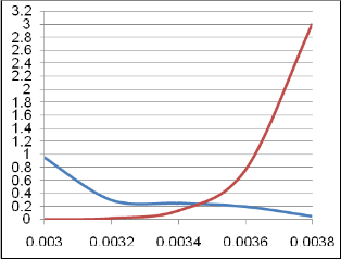
Fig.(6) The ROC curve for the local average of Veins Directions
Another performance parameter in the recog-nition system is the time. The time details of the best achieved recognition rate are shown in table (3).
In this work, we have proposed a reliable palm vein recognition and verification system. The proposed method to enhance the image and veins localization shows a high performance to extract the veins network, even though the vein images have poor-quality and suffer from many problem (blurry, noisy, etc.. ). Also, anew algorithm for feature extraction is proposed; it depends on the local average of veins direction.
IJSER © 2014 http://www.ijser.org
International Journal of Scientific & Engineering Research, Volume 5, Issue 4, April-2014 1033
ISSN 2229-5518
The experimental results show that our system achieved high recognition rate 99.95%, and EER =0.24% which indicate high per- formance in verification. The total recognition time is around
0.33ms; which is fast enough for real time applications.
At present, the proposed system can be applied to various parts of the human body where the veins are accessible (like: Finger, wrist, and etc). The quality of image data is vital for the application; hence more work is needed in the data preprocessing stage. So, the current image enhancement methods can be improved to provide better en- hancement results with lower complexity and time. Finally, attempts shall be made to integrate the vein pattern biometrics with other types of biometrics to become a multi-modal biometric system. And the candidate biometric technologies under current research include hand geometry and palm-print recognition.
[1] Rao, T. V., Preethi, K., “Future of Human Security Based on Computa- tional Intelligence Using Palm Vein Technology”, Global Journal of Computer Science and Technology, Vol. 10, Issue 10, Pp. 68-73, 2010.
[2] Wilson,C.,”Vein Pattern Recognition”, Taylor and Francis Group, 2010. [3] Lee, J.-C., “A novel biometric system based on palm vein image“, “Pat-
tern Recognition Letters”, Elsevier B.V., Pp. 1520–1528, 2012.
[4] Wang, J.-W., Chen, T.-H., “Building Palm Vein Capturing System for Extraction”, IEEE 21st International Conference on Systems Engineer- ing, 2011.
[5] Zhang, Y.-B., Li, Q., You, J., and Bhattacharya, P., “Palm Vein Extrac-
tion and Matching for Personal Authentication”, Springer-Verlag Ber- lin Heidelberg, Pp. 154-164, 2007.
[6] Heenaye- Mamode, M., Mamode, N. Ali., “A New Method to Extract Dorsal Hand Vein Patternusing Quadratic Inference Function”, Inter- national Journal of Computer Science and Information Security, Vol. 6, No. 3, 2009.
[7] Wang, Y., Li, K., Cui, J., ”Hand-dorsa Vein Recognition Based on Parti- tion Local Binary Pattern”, IEEE, 2010.
[8] Wang, Y.-D., Yan, Q.-Y., Li, K.-F., ”Hand Vein Recognition Based on
Multi-Scale LBP and Wavelet”, IEEE, 2011.
[9] Liu, J., Zhang, Y., “Palm-Dorsa Vein Recognition Based on Two- Dimensional Fisher Linear Discriminant”, IEEE, 2011.
[10] Wang, Y., Fan Y., Liao, W., Li, K., Shark, L.-K., Varley, M. R., “Hand
Vein Recognition Based On Multiple Keypoints Sets”, IEEE, 2012.
[11] Tang, Y., Huang, D., and Wang Y., ”Hand-dorsa Vein Recognition based on Multi-level Keypoint Detection and Local Feature Matching”,
21st International Conference on Pattern Recognition (ICPR),pp. 2837-
2840, 2012.
[12] Prabu, S.M., Sivanandam, S.N., “A Novel Biometric system for Person Recognition Using Palm vein Images”, International Journal on Com- puter Science and Engineering (IJCSE), Vol. 5, No. 08, Aug 2013.
[13] Nixon, M., and Aguado, A., “Feature Extraction & Image Processing for Computer Vision”, Third Edition, Academic Press,2012.
[14] Multispectral PolyU database, ww4.comp.polyu.edu.hk/~biometrics/.
[15] Zhang, H., Hu, D., “A Palm Vein Recognition System”, IEEE, 2010.
[16] Merouane, A., Benziane, S., Boulet, P., Benyamina, A. E.- H., Loukil, L,, “Hybridization of Discrete Binary Particle Swarm Optimization and Invariant Moments for Dorsal Hand Vein Feature Selection”, IEEE,
2013.
[17] Polli, R. M., Maran, A. V., Jouglas, A. T. Z, Silva E., Brandi, P. S., Hass D. I., “A proposal for the Hand Palm Identification, using Local Binary Pattern”, International Journal of Advanced Engineering Sciences and Technology (IJAEST), Vol. 9, Issue 2, Pp. 302 – 309, 2011.
IJSER © 2014 http://www.ijser.org