International Journal of Scientific & Engineering Research, Volume 5, Issue 7, July-2014 343
ISSN 2229-5518
“New Approach for Automatic Separation of ROI
and BG in Crime Scene Images and
Compression using DWT (ASRBDWT)”
Yogesh Dangar1, Vinita Shah2, Bhargesh Patel3
GCET, VallabhVidyanagar, Anand, Gujarat, India1, 2, 3 yogeshdangar202@gmail.com#1, shahvinita89@gmail.com#2, patel.bhargesh@gmail.com#3
Abstract-Crime sc ene imag es are very s ens itive to do any kind of preproc ess ing and c ompress ion, but the us e of images is increasing in expon ential mann er in crime detection and c rime s olving, s o we require to c ompress the crime sc en e images as well. For more c ompression ratio we c an us e Region of Interes t (ROI) c ompress ion. For c rime sc ene imag es our ROI may be evidenc es of crime. W e might have multipl e ROIs in c rime sc ene imag es. Som etimes it may n ot possible to s elec t ROI manu ally; bec aus e ROI may be too s mall and even s ometimes we c an miss s om e evidenc es in manual ROI s election. The s olution to this problem is autom atic s epar ation of ROI and bac kground (BG). In this paper, we had implem ented on e alg orithm f or autom atic s eparation of ROI and BG f or crime sc ene imag es. W e had us e c olor crime sc en e image f or automatic s eparation of ROI and BG and then c ompression is don e us ing DW T.
Key words: ROI, BG.
—————————— ——————————
separation of ROI and BG. By this method we can separate the
I. INTRODUCTION
In conventional compression model, an entire image is compressed with single compression ratio, i.e. equal or same level of compression is applied to the useful area as well as to the redundant area of an image. But in crime image compression it is desired to preserve the quality of a particular portion of an image more as compared to the rest of the image. The disadvantage of a conventional compression system is that it will compress the entire image with same compression ratio. Hence we cannot get a good overall compression performance in case of a conventional compression algorithm. And that is where a newer concept of compression, called the Contextual compression arises where the important and unimportant areas of an image are compressed with different compression ratios [1][3][4].
Now, crime scene images comprise of: Region of Interest (ROI), that used for the diagnosis and unimportant area- Background region, which comprises the less important information and is redundant. The background area in a crime image is quite large and we can compress it with quite a large compression ratio as it contains the redundant information. Again we cannot compress the diagnostically important area (ROI) beyond certain CR, in order to retain quality of the reconstructed image. Hence, the Contextual compression aims at compressing the ROI with the best quality (and least CR) and compressing the background with poor quality (and highest CR) to attain an overall better compression performance.
In crime scene images there are multiple ROIs and some of the ROIs are too small like bullets of gun, so we may miss to select that kind of ROIs in manual selection procedure. We had proposed one solution for this problem which is automatic
ROI and BG and then compress them separately.
The other use of this method is that if some evidences are too small and not found by detectives but they are present in an image then if we apply this method to that crime scene image then those evidences can be highlighted.
II. PROBLEMS IN AUTOMATIC SEPARATION OF ROI AND BG
The ROI is not fixed for all crime scene images. Even if the ROI is same then also the color and size of the ROI may vary. Automatic separation method is available for only two kinds of ultrasound kidney images (Ultrasound Transverse and longitudinal kidney images) [2]. There is no any general method exist for automatic separation of ROI and BG. In particular crime scene images, we have multiple, and some very small ROIs are there because of evidences. The most important reason is that the ROI is totally subjective matter.
III. PROPOSED SOLUTION FOR AUTOMATIC SEPARATION OF ROI AND BG
We can separate the ROI and BG automatically by performing edge detection operation and morphological closing operation. First we will convert the RGB image to gray scale image then we will apply canny edge detection technique to find edges and then we will apply the morphological closing operation to fill white color in the objects. Then we will separate the R, G and B component of an original color image, and subtract R, G and B component from the resulting gray scale image.
IJSER © 2014 http://www.ijser.org
International Journal of Scientific & Engineering Research, Volume 5, Issue 7, July-2014 344
ISSN 2229-5518
The advantage of automatic ROI selection is that it will help you in finding some evidences which are not visible in images normally but they exist. Because when automatic selection is done than only BG area will be selected and the color of BG will change so we can recognize the hidden evidences.
Following are the steps for ASRBDWT algorithm, Step 1) Read the image.
Step 2) Convert the RGB image to gray scale image.
Step 3) Apply canny edge detection technique to detect the edges on resulting gray scale image of step(2).
Step 4) Apply morphological closing operation on resulting image of step(3) for filling the objects with white color.
Step 5) Invert the white and black color of resulting image of step(4).
Step 6) Separate the R, G and B component of an original colored (RGB) image.
Step 7) Subtract the R, G and B component from the resulting image of step(5) image using.
Step 8) Combine the R, G and B component of step(7).
Step 9) Subtract resulting image of step(8) from the original image. By this step we will get separated background image.
Step 10) Apply forward DWT for encoding of background portion of an original image.
Step 11) Calculate the wavelet coefficients of BG.
Step 12) Quantize the wavelet coefficients for each subband of
BG.
Step 13) Get the compressed bit stream for BG.
Step 14) Combine the image of step(13) (Compressed BG image) and image of step(8) (ROI portion of an original image).
IV. RESULTS AND DISCUSSION
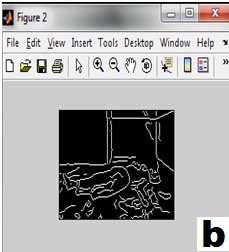
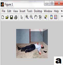
Figure 4.1 shows the results of proposed ASRBDWT algorithm; In the following image our region of interest is dead body, gun and the blood on the wall, and rest of the portion is consider as BG. Figure 4.1(a) represent the original image, Figure 4.1(b), shows the result of canny edge detection technique which we had applied to original image. Figure
4.1(c) shows the result of morphological processing applied on
result of canny edge detection. Figure 4.1(d) and (e) shows the separated ROI and BG portion respectively. Figure 4.1 (f) shows the resulting compressed image.
IJSER © 2014 http://www.ijser.org
International Journal of Scientific & Engineering Research, Volume 5, Issue 7, July-2014 345
ISSN 2229-5518
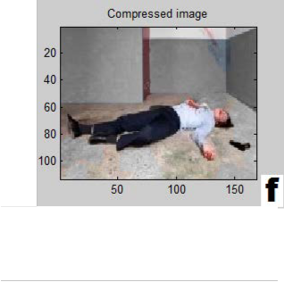
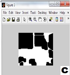
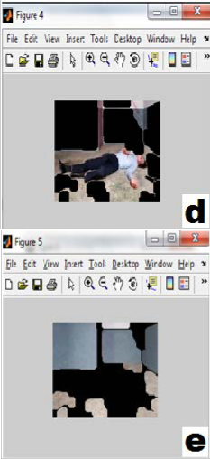
Fig. 4.1 (a)Original crime scene image (b)Canny edge detection (c)Morphological processing (d)Separated ROI portion (e)Separated BG portion (f)Compressed image
In this algorithm we are not applying compression of any kind to the ROI portion, because ROI portion in the crime scene images contains very sensitive information. The compression is applied only to the BG portion of the crime scene image. This way we will get good compression ratio as well as good image quality in ROI portion.
Table 4.1 shows the statistical analysis of ASRBDWT algorithm with parameter Bits per pixel, Compression ratio, Mean square error and Peak signal to noise ratio. We can see from the Table 4.1 that as the decomposition level increases the CR will increases and as CR increases, PSNR decreases.
Sr. No. | Decomposition level | BPP | CR | MSE | PSNR |
1 | 1 | 1.24 | 51.52 | 0.7132 | 49.60 |
2 | 2 | 1.24 | 84.44 | 6.2510 | 40.17 |
IJSER © 2014 http://www.ijser.org
International Journal of Scientific & Engineering Research, Volume 5, Issue 7, July-2014 346
ISSN 2229-5518
3 | 3 | 1.24 | 95.58 | 14.5032 | 36.52 |
4 | 4 | 1.24 | 98.80 | 27.5000 | 33.74 |
Table 4.1 Results of ASRBDWT algorithm
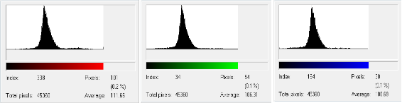
(a) Histogram of original image with R, G and B components
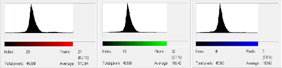
(b) Histogram of compressed image with R, G, and B components
Figure 4.2 Histograms of original and compressed image
[4] Sejal Thakkar and Yogesh N. Dangar,”Performance Analysis of
Figure 4.2(a) shows the histogram of original image and Figure
4.2(b) shows the histogram of compressed image with R, G
and B components.
V. CONCLUSION AND FUTURE WORK
The proposed ASRBDWT provides automatic separation of ROI and BG, and separate compression for BG using DWT compression technique. This algorithm provides good results for crime scene images with multiple ROIs.
Some other compression algorithm like JPEG2000, SPIHT etc. can be used with automatic separation of ROI and BG so that we can see more adequate results in the future.
REFERENCES
[1] Yogesh N. Dangar, Sejal Thakkar, “New Approach for Automatic Separation of ROI and BG in Crime Scene Images”, International Journal of Advancements in Research and Technology, Volume 2, issue 4, April 2013 (ISSN 2278-7763).
[2] Wan M. Hafizah, Eko supriyanto, “Automatic Generation of
Region of Interest for Kidney Ultrasound Images Using Texture Analysis”, International Journal of Biology and Biomedical Engineering, issue 1, volume 6, 2012.
[3] M.A. Ansari and R.S. Anand, “DWT Based Context Modeling of Medical Image Compression”, Department of Electrical Engineering, Indian Institute of Technology Roorkee, Roorkee-
247667 INDIA. E-mail: ma.ansari@ieee.org, XXXII NATIONAL SYSTEMS CONFERENCE, NSC 2008, December 17-19, 2008.
Crime Images Using CROI with JPEG WAVELET”,International
Journal of Scientific and Engineering Research, Vol. 4 March 2013
[5] R. K. Kher, C. K. Modi and R. S. Anand, “Ultrasound Medical Image Compression Using Contextual Approach”, International Conference on Recent Advancements and Applications of Computer in Electrical Engineering (RACE 2007) at Engineering College, Bikaner during March 24-25, 2007.
[6] M. A. Ansari and R. S. Anand, “Performance Analysis of Medical
Image Compression Techniques with respect to the quality of compression”, Published in IET-UK International Conference on Information and Communication Technology in electrical sciences (ICTES 2007), Dr. M.G.R. University, Chennai, Tamilnadu, India. Dec. 20-22, 2007. pp. 743-750.
[7] Rafael C. Gonzalez and Richard E. woods, “Digital Image
Processing”.
First Author: Mr. Yogesh N. Dangar had completed his M.E. in Information Technology from Gujarat Technological University in year 2013. He is working as Assistant professor in department of information technology at G. H. Patel College of Engineering and Technology, V.V. Nagar.
Co-Author: Ms. Vinita Shah had completed her M.E. in Information
Technology from Gujarat Technological University in year 2013. She is working as Assistant professor in department of information technology at G. H. Patel College of Engineering and Technology, V.V. Nagar.
Co-Author: Mr. Bhargesh Patel had completed his M.Tech. in Computer Engineering from Dharmsinh Desai Univeristy, Nadiad in year 2011. He is working as Assistant professor in department of information technology at G. H. Patel College of Engineering and Technology, V.V. Nagar.
IJSER © 2014 http://www.ijser.org
International Journal of Scientific & Engineering Research, Volume 5, Issue 7, July-2014
ISSN 2229-5518
347
I£ER 2014 http://WWW.ISer.org