International Journal of Scientific & Engineering Research, Volume 4, Issue 12, December-2013 2232
ISSN 2229-5518
Fluorescence Spectrum of Alocasia culcullata Schot and Chlorophyll Laser
1Mitali Konwar and 2G.D.Baruah
1Digboi Mahila Mahavidalaya,Digboi-Tinsukia-786171(India)
2Centre for Laser &Optical Science, New Uchamati,Doomdooma-786151 (India) Email:mitalikonwar@rediffmail.com
Abstract
The present work reports the fluorescence spectra of the acetone extract of some medicinal plant leaves and specifically the fluorescence spectrum of the acetone extract of the plant leaves Alocasia Culcullata Schot (local name “kolia kosu”) has been studied in detail due to its importance in connection with its potential as dye laser. It is worthwhile to note here that the acetone extract of the plant leaves of this particular specimen exhibits very intense fluorescence when it is excited with the help of a radiation from a broadband source of 500 watt halogen lamp or from a 30 mW green diode laser. We have studied the fluorescence by placing the system inside a laser cavity of length 12cm and studied the characteristic fluorescence as it emerges out of the cavity. The fluorescence is accompanied by gain. We term it as chlorophyll laser.
Keyword: Chlorophyll laser.
.
1. Introduction
Nearly all vegetations from the tiny grasses to the lofty trees in the forest exhibit green as its
natural color to be extracted easily. The cuvette is then held against a bright source of light which be broad or monochromatic. The spectrum of light coming from the cuvette and analyzed by the spectrometer as absorption or fluorescence radiation
IJSER
prominent color which is undoubtedly a great
environmental phenomenon on the earth. It is worthwhile to note here that Sir C.V. Raman [1] was particularly concerned with the studies related to the green color exhibited by plant leaves and the colors exhibited by foliage and flowers. These are the colors which are actually perceived and it is their relationship to the spectral character of the light reaching our eyes which was the subject of study in case of Sir C.V.Raman. Raman describes in a most elegant and simplified manner the physical process involved. Sunlight is incident on the leaves of growing vegetation or on the petals of flowers. It enters the material and re-emerges after internal reflections (or diffusions) or scattering. It may also be accompanied by light which is reflected or diffused at the surface of the leaves or petals. They are usually not important and their effect may be minimized by an appropriate choice of the direction of observation. They may be completely avoided if the light which emerges after passing through the leaves or petals is examined through a pocket spectroscope. The regions of the spectrum of which there is a strong absorption (black region) or emission (colored region) may be identified. This is presumably a rough estimate but the work may be supported by observation through a photometric spectrometer. What may be observed with absorption is also true for fluorescence, which is the main topic of discussion is the present work. In many cases immersion of the crushed plant leaves or petals in a cuvette containing a suitable solvent such as acetone enables the pigments responsible for their
can be examined. Extensive studies carried out by the
author [2] during recent years have enabled a comprehensive view to be obtained of the nature of the fluorescence or absorption spectrum and their relationship to the absorptive or fluorescent properties of the pigments contained in the material of the leaves or petals. We are concerned in the present work with the fluorescence spectra of few plant leaves of medicinal importance and specifically we shall describe the strong fluorescence from a sample of Alocasia Culcullata Schot [local name “kolia kasu”]and the possible realization of chlorophyll laser. Such type of strong fluorescence has not been reported earlier. We use the term chlorophyll laser to emphasize the fact that all the fluorescence originated from green plant leaves are due to chlorophyll molecule.
2. Experimental
We have considered seven specimens of medicinal plant leaves for our study and the fluorescence spectra of all these specimens shall be discussed in the present work along with the fluorescence spectrum of a specimen of Alocasia Culcullata Schot (local name “kolia kasu”). We have used a broad band source (500 mW Ar+ laser) for our work. It may be noted that for exciting the fluorescence a 500 Watt halogen lamp or a 30 mW green diode laser are also adequate for the purpose. The acetone extract of the specimens is kept in a cuvette of suitable dimension (1cm x 1cm x3cm) and
IJSER © 2013 http://www.ijser.org
International Journal of Scientific & Engineering Research, Volume 4, Issue 12, December-2013 2233
ISSN 2229-5518
the cuvette with the solution is held in front of the slit
of a glass spectrograph (ASCO two prism) having good dispersion in the visible region (3900-7000)A°. The radiation from the source is allowed to be incident on the sample cell with the help of a short focused convex lens and the fluorescence radiation is observed in the direction perpendicular to the direction of the incident beam.The fluorescence spectrum is recorded photographically with the help of a color film (400 ASA) which is commercially available. It is very easy to measure the intensity of the fluorescence bands with the help of software called “Image J”. This procedure makes the entire system a complete instrument and as good as any
recording spectrometer and in some cases even better [2]. The fluorescence intensity is estimated with the help of a silicon detector equipped with a digital multimeter (model PD 10 OPHIR). One can also observe the fluorescence radiation from the cuvette with help of a pocket spectroscope and measure the intensity of the red sector of the spectrum visually. It may be noted that the silicon photodiode is connected with an optical fiber which is kept in contact with the sample cell.
The experimental set up needed for the
Fig. 1 Experimental arrangement to observe LIF and possible gain from Alocasia Culcullata Schot.
3. Results and Discussion
It is now appropriate to produce the results in a comprehensive manner. The acetone extract of the plant leaves alocasia Culcullata Schot exhibits very intense fluorescence when the radiation from a source of halogen lamp or a DPSS green laser is incident on the sample cuvette. It is worthwhile to note that dark room is illuminated with this red fluorescence similar
IJSER
study of modification of the fluorescence and
possible laser action is quite simple. The cuvette with strong fluorescence from the specimen of Alocasia Culcullata Schot is placed between two dielectric mirrors (obtained from a He-Ne laser tube) separated by a distance of 12 cm. The mirrors are placed on precision mounts suitable for 1" optics with micrometer controlled stages for X-Y-Z tilt. For proper alignment it is essential to view the fluorescence visually along the direction perpendicular to the planes of the mirrors. By trial and error methods it has been observed that at a particular position of the mirrors the intensity of radiation emerging through the mirrors suddenly becomes very high. A sheet of ground glass interposed between the mirror and the eye helps to visualize this effect. We term this position where a bright red spot is observed as position X. It is worthwhile to note here that the bright spot is seen on the ground glass even at a distance of 50cm from the mirror. The observations should be made in a completely darkened room. The functional block diagram of the experimental arrangement for LIF and to observe possible gain is shown in Fig. 1.
to the red light from a 15 Watt bulb. Fig. 2 shows the
projected image of the light at a distance of 1 metre from the sample cell.
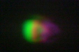
Fig.2 Projected image of the red fluorescence
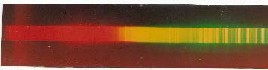
This type of strong fluorescence is not seen in any of the fluorescence that we have examined for acetone extracts of other plant leaves. When photographed on a glass spectrograph the fluorescence spectrum exhibits a broad and intense band in the red sector ranging from 6300 to 6900 A°. Fig. 3 shows the fluorescence spectrum along with the intensity distribution curve worked out with the help of the software (image J).
IJSER © 2013 http://www.ijser.org
International Journal of Scientific & Engineering Research, Volume 4, Issue 12, December-2013 2234
ISSN 2229-5518
Table 2. Pocket spectroscopic observation of the intensity of red fluorescence
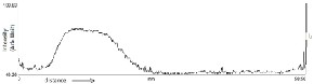
Fig 3: a) Fluorescence spectrum of Alocasia
Culcullata Schot
b) Intensity distribution curve
The characteristic feature of this spectrum is the absence of the green sector which is presumably extremely weak.
At this stage it is worthwhile to make a comparative estimate of the fluorescence intensities of radiations from the acetone extracts of the plant
w= weak, ms=medium strong, vw= very weak, vs=
very strong
4. Cavity Modified Fluorescence
We shall concern ourselves with the following problems. How sensitive are the fluorescence when they are kept inside a cavity as described in Fig 1. What are the factors which results in the modification
IJSER
leaves measured with the help of photodiodes
connected with optical fiber. The results are shown
below in Table 1.
Table 1. Relative intensities in the red sector of the radiations measured with photodiodes.
Specimens | Name | Intensity |
1 2 3 4 5 6 7 ** | Alocasia Culcullata Schot Mesua ferra –L Metha Spicata Linm Azadirachta indica Lawsonia syn Cassia fistula Linn Artemisia vulgaris Linn --------------- | 1455 20 35 10 5 50 16 1500 |
We also include here the pocket spectroscopic observations of the intensities in the red sector of the spectrum. The results are shown in Table 2. The findings in Table 2 are confirmed by the measurements of the photodiode meter readings of the intensities of radiations of different samples. As may be inferred from these measurements the intensity of the fluorescence radiation originating from Alocasia Culcullata Schot is unbelievably strong. Similar is not the case for other specimens.
of the fluorescence inside a cavity and also possible

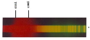
gain. It is apparent that these problems can only be solved by systematic observational studies, though it is possible to venture on some general considerations based on the theory of dye laser. As shown in Fig 1 the radiation from the fluorescence with maximum intensity (marked as X) observed on the ground glass is allowed to be incident on the slit of the spectrograph with the help of a short focused lens. The resulting spectrum is recorded on a commercially available film (400 ASA). The spectrum is reproduced in Fig 4 along with the spectrum recorded without the use of the cavity. The brightness of the radiation spot (marked as X in Fig 1) as actually observed on the ground glass can also be measured with the help of the photodiode connected by an optical fiber.
IJSER © 2013 http://www.ijser.org
International Journal of Scientific & Engineering Research, Volume 4, Issue 12, December-2013 2235
ISSN 2229-5518
Fig 4 a) Fluorescence spectrum of the acetone extract of Alocasia Culcullata Schot without cavity
b) Spectrum with cavity
As may be inferred from Fig 4b, highly remarkable and significant changes are observed in the fluorescence spectrum when it is put inside a cavity. The fluorescence is modified and narrowed down to a considerable extent The broad band in the red sector from 6300-6900 A° is completely absent but a narrow but strong line appears at 6830 A°. It follows from the foregoing observations that the fluorescence spectrum of the specimen under consideration apparently undergoes modification, due to the introduction of the cavity. These changes are not observed in the case of fluorescence of other specimens, that the changes observed in the fluorescence spectrum of the acetone extract of the
specimen is due to the introduction of the cavity is
Fig 5 Chlorophyll –a molecule
absorbing light which excites the electrons within the molecule and this produces a series of complex reactions. From the absorbing characteristics or absorption spectra of chlorophyll a and b it is obvious that chlorophyll is fantastic at absorbing violet, blue and red light but poor absorber in green, yellow and orange light. It may be noted that the sun shines by
IJSER
obvious by the fact that a slight change in the alignment of the mirrors destroys the spot. This can be observed visually. Observations by the photographic technique confirm the remarkable finding that the spectra observed in the red sector range may be either cavity modified or gain due to ASE. It appears appropriate to conclude the present work with some comments on the origin of the fluorescence with its subsequent modification by cavity. As has already remarked in the beginning, the fluorescence is due to the molecule chlorophyll. The excitation of fluorescence with the help of laser radiation has become a procedure that has attracted research workers from various disciplines. LIF of green plants detection of vegetation stress has been initially proposed by Chapelle [3]. Later these studies have been used to explore the possibility of using laser as remote means of measuring vegetation characteristics such as plant vigour, natural mineral deficiency, plant type identification and biomass estimation [4-10]. Chlorophyll is a family of photoreceptive molecule by cynobacteria, algae and plants to perform photosynthesis. As may be inferred from Fig 5 this molecule is largely made of carbon and hydrogen and some nitrogen- containing molecules surrounding magnesium atom. There are several kinds of chlorophyll, prominent one is chlorophyll- a. chlorophyll functions by
far the brightest in the yellow sector of the spectrum,
the region where the chlorophyll absorption is minimum.
5. Conclusion
From what has been discussed above it is worthwhile to make an appropriate conclusion and give an outlook for any future work. The present work reports the fluorescence spectra of the acetone extracts of some medicinal plant leaves with reference to the fluorescence spectrum of a particular specimen called Alocasia Culcullata Schot where the fluorescence intensity is unusually strong. This characteristic has been investigated by inserting the cuvette inside a cavity. Modification of fluorescence takes place and gain is observed.
Reference :
1. 1. C.V.Raman, Proc. Indian Acad Sci A62 (1965).73-77
2. Mitali Konwar, “Optical and Spectroscopic studies of some medicinal plants and natural dyes" Ph.D. thesis, Dibrugarh university (2007), unpublished.
3. E.W. Chappell, F.M. Wood, W.W.
Newcomb and J.E.McMurthy, Appl. Opl. 23
(1985) 74-85
IJSER © 2013 http://www.ijser.org
International Journal of Scientific & Engineering Research, Volume 4, Issue 12, December-2013 2236
ISSN 2229-5518
4. H.K. Lichtenthaler and U.Rinderle CRC Critical Reviews in Analytical Chemistry 19 (1988).
5. R. Gopal, K.B.Mishra, M. Zeesham, S.M Prasad and M.M. Joshi, Curr.Sci. 83(7) (2002) 880-884
6. R.Valentini, G. Cecchi, P.Mazzingli, G.S.
Agati, M.Bazzani, P.De Angelis, F. Fusi,
G.Matteucci and V. Raimonli. Remote Sens. Environ. 47 (1994) 29-35.
7. Y.Saito, K.Hataka, E.Nomura, TD.
Kawahara and A. Nomura. Appl. Opt. 37
(1998) 437
8. G.A. Johanson, S.V. Mantha and T.A.Day
J.Plant. Physiol. 156 (2000) 242-252.
9. E.W. Chappell, J.E. McMurthy, F.M. Wood and W.W. Newcomb, Appl. Opl. 23 (1993)
139-142
10.M. Broglia: Appl.Opt. 32 (1993) 334-338.
IJSER
IJSER © 2013 http://www.ijser.org