The research paper published by IJSER journal is about Detection of Diabetic Retinopathy Using Sobel edge detection method in DIP 1
ISSN 2229-5518
Detection of Diabetic Retinopathy Using
Sobel edge detection method in DIP
Jyoti Patil
Assistant Professor
I2IT, Hinjewadi, Pune-411057 India
Email: jyot.physics@gmail.com
Dr. A. L. Chaudhari
H.O.D. Department of Electronics
MGSM’s Arts, Science & Commerce College,
Chopda Dist. Jalgaon 425 107 India
Email: chaudharial@yahoo.co.uk
Abstract— Diabetic retinopathy, a complication of diabetes that occurs as a result of vascular changes in the retina, It is a major cau se of loss of vision. Automated image processing has the potential to assist in the early detection of diabetes, by detecting changes in blood vessel patterns in the retina. Image processing techniques can reduce the work of ophthalmologists and the tools used automatically locate the exudates. 0In this paper the process and knowledge of Digital Image Processing (DIP) is used. Automated analysis techniques for retinal images have been an important area of research for developing screening programmers. By using MATLAB for programming to devel op the DIP tool for diagnosis of eye infection . Sobel edge detection algorithm is a method to find the edge pixels in an image. Edg es are pixels which carry important information in an image. Thus sobel method is best technique for features are extended & u sed to classify the pixels in the patch into vessel and non vessel.
Keywords— Diabetic Retinopathy, DIP, MATLAB,
1. INTRODUCTION
Diabetic retinopathy (DR) is a severe eye disease that affects many diabetic patients. Diabetic retinopathy is the most common cause of blindness which a complication of diabetes mellitus, so it is necessary to diagnosed early. The eye, a vital organ of the human body, gives us the sense of color, shape and state of physical objects. But if Abnormalities occurs in the eye because of diseases such as Conjunctivitis, Fungal Keratitis, glaucoma, diabetic retinopathy, fungal infection, diabetes then eye may be damaged [1].
The complicated images obtained from infected eye
will be processed using digital Image Processing (DIP)
technique, which manipulates the image for the purpose
of either extracting information from the image or produces an alternative representation of the image. Thus screening is the most effective method to detect early signs of diabetic retinopathy [2-3]. Using screening method big blood clots called hemorrhages, Hard exudates, The bright circular region from the blood vessels called optic disk, The fovea defines the center of the retina, and is the region of highest visual acuity, exudates and microaneurysms, irregular shaped, and found in the posterior pole of the fundus can be detected.
Ma et al. [3] defined a quality descriptor according to
three classes, namely, out-of-focus images, motion blurred images and severely occluded images of eyelids and eyelashes. Zhu et al. [4] proposed a quantitative quality measure using discrete wavelet decomposition.
Analyzing and interpreting retinal images have
become a necessary and important diagnostic procedure in ophthalmology. We are interested in vessel segmentation in color images for screening of diabetic
retinopathy. Thus to remove noise, enhance objects of interest - blood vessels, damaged areas, Changes in the
blood Vessel Structure we can use Sobel algorithm, and Laplacian of Gaussian operator, which detects the edges of blood vessels [5]. Micro aneurysms [tiny dilations of the blood vessels] are the first apparent sign of diabetic retinopathy so that their detection in fundus images through photography might be detect the disease in an early stage.
2. METHODS OF DETECTION
SOBEL EDGE DETECTION METHOD
Detection of vessels, exudates, and hemorrhages, blood clots, Hard exudates, optic disk is possible using Sobel method. Edge detection is the process of localizing pixel intensity transitions. The Sobel operator is an algorithm for edge detection in images discovers the boundaries between regions also it determine and separate objects from background in an image. It’s an important part of detecting features and objects in an image [6].
The Sobel method finds edges using the Sobel approximation to the derivative. It returns edges at
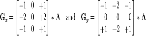
those points where the gradient of I is maximum. Where the gradient of the considered image is maximum. The horizontal and vertical gradient matrices whose dimensions are 3 × 3 for the Sobel method has been generally used in the edge detection operations [7]. If we define A as the source image, and Gx and Gy are two images which at each point contain the horizontal and vertical derivative approximations, the computations are as follows.
IJSER © 2012
http://www.ijser.org
The research paper published by IJSER journal is about Detection of Diabetic Retinopathy Using Sobel edge detection method in DIP 2
ISSN 2229-5518
Further gray scale image is processed, sobel method is applied to find out derivative of grayscale image therefore Marr Edges, sigma=1, Marr Edges, sigma=2 is applied. in Fig.3.A) & B) we can clearly observe separate edges of nerve fibers, clotted spots, hemorrhages, Hard
Exudates.
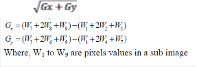
G= ; Where, |G|= |Gx|+|Gy| &
[7]. These filters estimate the gradients in the
horizontal (x) and vertical (y) directions and the
magnitude of the gradient is simply the sum of these 2
gradients. Using this information, we can also calculate the gradient's direction [8].Where, for example, Θ is 0 for a vertical edge which is darker on the right side.
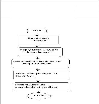
Fig. 1 Flowchart for Sobel Method
3 .RESULT & DISCUSSION
The Sobel method finds edges using the Sobel approximation to the derivative. The Sobel operator calculates the approximate image gradient of each pixel by convolving the image with a pair of 3×3 filters. Thus fig.2) a) is a original infected image of in which red spot shoes clotting of blood also yellowish spots shows hemorrhages, Hard exudates. This is then converted in to grayscale image [9].
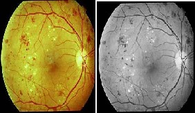
Fig.2. a) Pink Eye b) Gray Scale Imag
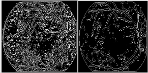
Fig. 3 A) Marr Edges, sigma=1 B) Marr Edges, sigma=2
Sobel method is able to detect exact location of optic disk with damaged blood vassals. We can observe yellowish part of optic disc in fig 3 a) which is damaged due to increase in pressure in eye due to Diabetic retinopathy which causes in blindness .So if it is possible to detect in early stage with the help of sobel function consist Marr Filter we can observe damaged part of optic disc, with damaged blood vassals, also we can observe yellowish blood spot due to pressure in eye. In Fig.b) & C).
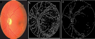
A) Original Diabetic image, B) Detected edges, C) Detection of optic disc. Effect application of Edge detection algorithms of sobal edge detection method on diabetic images using function of Marr Filter.
4. Conclusions
We proposed an efficient method based on Sobel method which differentiates between original diabetic image & processed image. Sobel method can show separates parts of the edges of nerves from whole image. This paper has demonstrated an automated system which is able to distinguish normal and abnormal vasculature on the optic disc. The main focus of this work is on segmenting the diabetic retinopathy image and classifies the Exudates, micro aneurysms and hemorrhages. These methods give almost good results. Thus Image processing techniques can reduce the work of ophthalmologists and the tools used automatically locate the exudates.
IJSER © 2012
http://www.ijser.org
International Journal of Scientific & Engineering Research Volume 3, Issue 7, July-2012 3
ISSN 2229-5518
ACKNOWLEDGMENT
I express my thanks to Dr.A.D.Shaligram, Head of Department of Electronics, University of Pune, for sharing his valuable knowledge.
IJSER © 2012 http /fwww l[ser org
The research paper published by IJSER journal is about Detection of Diabetic Retinopathy Using Sobel edge detection method in DIP 4
ISSN 2229-5518
REFERENCES
[1] Bill Silver, “An Introduction to Digital Image Processing”,
Cognex Corporation, Modular Vision Systems Division,2000.
[2] Mr. R. Vijayamadheswaran, Dr.M.Arthanari, Mr.M.Sivakumar,”
DETECTION OF DIABETIC RETINOPATHY USING
RADIAL BASIS FUNCTION”, Doctoral Research Scholar,Anna
University, Coimbatore.
Ma, L., Tan, T., Wang, Y., Zhang, D.: “Personal Recognition
Based on Iris Texture Analysis”, IEEE Trans. Pattern Analysis
and Machine Intelligence,25(12) (2003) 1519–1533.
[3] Zhu, X., Liu, Y., Ming, X., Cui, Q.: “A Quality Evaluation
Method of Iris Images Sequence Based on Wavelet Coeffi-cients in „Region of Interest‟”, Proc. of the 4th Int. Conf. on Computer and Information Technology, pp.24 -27, 2004.
[4] Big Fang,Wynne, HsuMong Li Lee‟” On the Detection of Retinal Vessels in Fundus Images” , Singapore-MIT Alliance , National University of Singapore, Department of Computer Science, School of Computing National University of Singapore.
[5] Iqbal, M.I , Aibinu, A.M , Gubbal, N.S . Khan, “AUTOMATIC
DIAGNOSIS OF DIABETIC RETINOPATHY USING FUNDUS
IMAGES”, Master‟s Thesis Blekinge Institute of Technology
October 2006
[7] http://en.wikipedia.org/wiki/Sobel_operator
[8] www.utdallas.edu/~dxa081000/IMAGEFILTERING.ppt
[9] V.Vijaya Kumari,” Diabetic Retinopathy-Early Detection Using
Image Processing Techniques”, Department of ECE, V.L.B.
Janakiammal College of Engineering and Technology Coimbatore
641 042, India.
Name of Author: Ms.Jyoti Devidas Patil
Born in 1983, obtained M. Sc., M. Phil degrees from
North Maharashtra University, Jalgaon.Joined the
Department of Physical Science.Presently Working as
Assistant Professor in Engineering Science Department of “International Institute Of Information Technology, Pune” Currently working on “Digital Image Processing”
Papers published :
1)international conference proceedings, “Proceedings of
the DRDO sponsored eighth Control Instrumentation System Conference CISCON-2011 (An International Conference) November 3-6,2011, Karnataka, Manipal University, Manipal,” (Development of Digital Image Processing tool for diagnosis of eye infection using MATLAB)
2) International Workshop On Green Energy
Technologies (IWGET),(Production of biodiesel using

jatropha).
Name of Guide - Dr. A. L. Chaudhari
Dr. A. L. Chaudhari, born in 1964, obtained M. Sc.,
M. Phil. & Ph. D. degrees from University of Pune.
Joined the Department of Electronics, Arts, Science & Commence college, Chopda Dist. Jalgaon, India as lecturer since 1988 & presently working as Associate Professor, Head of Electronics & Computer science department. He is guiding students for M. Phil. & Ph.D.Co-authored 10 books & published 25 papers in national & international journals/conference proceedings. Visited National university of Singapore
& University of Tokushima, Japan. He is executive council member of IETE(Pune Chapter),member of
Board of Studies & member of Faculty of Science, North Maharashtra University, Jalgaon Completed minor research project sponsored by UGC. His current area of research is fiber optic sensor, PC based instrumentation & image processing.
IJSER © 2012
http://www.ijser.org
n
e
n a ti
0 n a
0
u
n
a
1
0
f
s
c
e n ti fi
c
&
E n p
1
n e e
n
g R e
s
e
IJ
s
E R
©
0
b.
ll
2.
.!.

n
e
n a ti
0 n a
0
u
n
a
1
0
f
s
c
e n ti fi
c
&
E n p
1
n e e
n
g R e
s
e
IJ
s
E R
©
0
b.
ll
2.
.!.
