International Journal of Scientific & Engineering Research, Volume 5, Issue 7, July-2014 1167
ISSN 2229-5518
Computation of Brain Asymmetry in 2D MR Brain
Images
P. Kalavathi
Gandhigram Rural Institute – Deemed University, Gandhigram, Tamil Nadu, India
Abstract—The automatic computation of brain asymmetry is needed to study structural, volumetric and functional differences be- tween the left and right brain structures to quantify and correct several MR brain deformities. This paper proposed a method to compute brain asymmetric measures such as Asymmetric Volume Index (AVI) and Asymmetric Shape Index (ASI) between the segmented left, and mirrored and registered right brain images. In order to register the right brain with the left brain for shape asymmetric measure, an image registration method based on Fourier-Mellin (FM) transformation is developed as a part of this brain asymmetric analysis.
Index Terms— MRI brain structure, brain asymmetric analysis, MRI brain images, asymmetric volume index, asymmetric shape index
1 INTRODUCTION
—————————— ——————————
HE The human brain exhibits an approximately bilateral symmetry across the sagittal plane. A longitudinal fissure
cussion are given in section 3 and the conclusions is given in section 4.
separates the human brain into two distinct cerebral hem-
ispheres which divides the brain into two equal parts. Howev-
er, these two parts are almost never perfectly symmetric even for the normal brains [1][2][3].
Brain morphometric studies often incorporate compara- tive asymmetric analyses of the left and right hemispheric brain structures. Brain asymmetry is thought to originate from evolutionary, developmental, hereditary, experimental and pathological factors and it has been correlated with asymmet- rical behavioral traits such as handedness, auditory percep- tion, motor preferences and sensory acuity [4]. Moreover, brain asymmetry analysis provides methods for computer assisted diagnosis for mental diseases such as schizophrenia [5][6][7]. Several methods have been proposed for brain seg- mentation [9]-[12], volumetric and structural analysis of brain structures based on interhemisphere asymmetric [13][14][15], asymmetric analysis using voxel-based morphometry [16][17], surface based approaches for asymmetric study[18][19] and asymmetry computation in terms of regional tissue composi- tion [20][21].
In this paper, a new automatic method to compute asymmetric measures such as Asymmetric Volume Index (AVI) and Asymmetric Shape Index (ASI) are calculated be- tween the segmented left and right brain structures. To com- pute ASI, the mirrored right brain structure is need to be regis- tered with the coordinate space of left brain structure. For this purpose, an image registration method based on Fourier- Mellin Transformation (FMT) [22] is developed. The proposed method was tested with 18 volumes of T1-weighted brain im- ages obtained from Internet Brain Segmentation Repository (IBSR) [23] which includes the delineated brain volumes of all these brain volumes. Computed AVI and ASI values show that the proposed method accurately calculated the asymmet- ric measures in MR brain images. The remaining part of the paper is organized as follows: In section 2, the methodological details of this proposed method is given. The results and dis-
Fig. 1. Flowchart of the proposed method.
IJSER © 2014 http://www.ijser.org
International Journal of Scientific & Engineering Research, Volume 5, Issue 7, July-2014 1168
ISSN 2229-5518
2 METHODS AND MATERIALS
2.1 Brain Asymmetric Analysis
The brain asymmetry analysis is necessary to compute the anatomical and volumetric difference between the left and right brain structures. Even the left and right brain with equal volume may have different shapes. Therefore, a detailed anal- ysis is needed to understand the human brain and its changes for accurate diagnosis of various brain related diseases. The flowchart of the proposed method is depicted in Fig. 1. The proposed method first reads the input brain image I and its corresponding delineated brain image X. In the selected brain images dataset each volume is attached with its delineated image volume containing labeled brain image for all its brain structures. The proposed method reads only the left and right hemispheres in the delineated image and store it as image L and image R respectively. These images are quantitatively
analyzed to measure the volumetric difference by calculating
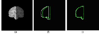
Fig. 2. Shape comparison of left and right brain structures; (a) Segmented left and right brain structure (b) Contours of left (white) and mirrored right brain (green) structures (c) Contours of left (white), and mirrored and registered right brain (green) structures
First the input images are converted into Discrete Fourier Transformation (DFT). The DFT of an image f(x,y) of dimen- sion M×N, where, x=0,1,2,…,M-1 and y=0,1,2,…,N-1 is given by:
M −1 N −1
AVI [23] measure by applying the following equation.
L − R

F{f(x,y)}=F(u,v)= 1
MN
∑ ∑ f ( x, y )e − j 2 π( ux / M +vy / N ) (3)
x =0 y =0

AVI = 2
L + R
(1)
where u=0,1,2,…,M-1, v=0,1,2,…,N-1 and j=

− 1 .
where, L represents the total pixels in the left brain and R
represents the total pixels in the mirrored right brain
Then, the inverse of DFT is given by:
Then the quantitative shape difference is measured as per the equation (2).
F′{F(u,v)}=f(x,y)=
M −1 N −1

∑ ∑ F ( u ,v )e j 2 π( ux / M + vy / N )
MN x = 0 y = 0
(4)
2( L ∩
ASI = 1 −
R1 )
Then the Log-Polar Transformation (LPT) is applied on the
 L
L +
+  R1
R1 
(2)
transformed images. An LPT is a non-linear and non-uniform sampling of the spatial domain. In the log-polar (log r, θ) co-
where, L represents the total pixels in the left brain, and R1
represents total pixels in the mirrored and registered right
brain. The deviation of AVI and ASI measures from zero is
analyzed and normally it ranges from -1 to +1.
ordinate system, r denotes radial distance from the center ( xc , yc ) and θ denotes the angle of rotation. Hence, any arbi- trary point (x,y) chosen from an image can be expressed in the form of polar coordinates as:
In order to study shape the asymmetric bias, the right brain image R is mirrored to produce the mirrored image MR and then the image MR is registered against the left brain im-
r = log

base (
( x − xc
)2 + ( y − y
)2 )
(5)
age L. It is necessary to register the image R with the image L,
y − y
because the mirrored right brain images may not always get aligned with the same coordinate space of left brain as repre-
φ = tan −1 c
x − xc
(6)
sented in Fig. 2. Therefore, the mirrored right brain image R
has to be registered to the coordinate space of the left brain L
prior to the computation of ASI.
Image registration is a method to align the reference im- age in the same geometrical space of base image. Fourier- based methods are the efficient and accurate method to esti- mate the image transformation such as rotation, scaling and translation for image registration [24]. These methods search for an optimal match for the images as per the information in the frequency domain. This proposed method uses Fourier transformation, log-polar transformation and phase correla- tion methods.
Applying a polar coordinate transformation to an image, maps the lines in Cartesian space to the horizontal lines in the polar coordinates. In this method, the logarithmic conversion to obtain the polar coordinates uses base 10. Then it applys DFT on the polar transformed images to compute phase corre- lation. The Fourier magnitude in polar coordinates differs only by translation. The phase-correlation method is used to find this translation. Phase correlation is a method of image regis- tration and uses Fast Fourier Domain approach to estimate the relative translation between two images. Correlating the mag- nitude of a FMT, it is possible to obtain an image registration method invariant to translation, rotation and scaling. Then the
IJSER © 2014 http://www.ijser.org
International Journal of Scientific & Engineering Research, Volume 5, Issue 7, July-2014 1169
ISSN 2229-5518
scale and rotation parameters are derived by calculating the cross-power spectrum. The cross-power spectrum (R) of two
B1 .
images f and defined as:
F F 1*
f 1 with Fourier transforms F and F1 is


R = F1 1*
F F
(7)
2.2 Brain Image Datasets Used
Eighteen volumes of T1-weighted images were obtained from the IBSR of the Centre for Morphometric Analysis
where, F1* is a complex conjugate of F1, the phase of the cross-power spectrum is equivalent to the phase difference between the images. Then, the rotation ∆x and the scale ∆y is computed by:
(CMA) at the Massachusetts General Hospital. Each volume consists of 128 two-dimensional sequential coronal slices with
dimensions of 256×256 pixels and the slice thickness is 1.5mm. These MRI scans are acquired from all age groups including
juvenile to old age. The IBSR also maintains the manually
(∆x,∆y) = arg max{R}
( x ,y )
(8)
segmented (ground truth or gold standard) brain mask and delineation of the brain structures performed by trained ex-
where, (x,y) is the location of the peak in R. After computing the rotation and scaling parameters, the referenced image is rotated and scaled accordingly to register with the base image. Again it applies DFD on the rotated image and computes the phase correlation to obtain the shift parameters for translation.
The summary of steps involved in the proposed image registration method are described in Alg. 1.

Alg. 1. Image Registration
perts. Several volumes of these dataset had relatively low con- trast images.
3 Results and Discussion
This method is applied on all the volumes of the selected dataset and found that the proposed method have accurately computed the asymmetric bias on all the images. To explore the efficiency of this method on asymmetric analysis, a set of sample images and their corresponding left brain, right brain, overlapping the contours of left and mirrored right brains be-
fore registration and after registration are shown in Fig. 3.
Input : Base image A and Reference image B.
Output: Registered image B1 .
1. Apply DFT on the input images A and B and shift its zero-frequency component to the center of spectrum and obtain FA and FB.
2. Perform LPT to transform FA and FB into log- polar space to obtain the image LA and LB .
3. Apply DFT on LA and LB to get QA and QB and compute the phase correlation of QA and QB to obtain r.
4. Find the location (x,y) in r of the peak of the phase correlation.
5. Compute angle of rotation θ = (360/size(r)) ×
y and rotate the image B by -θ to get BR .
For the segmented left brain Fig. 3(b), right brain Fig. 3(c) and the outer contours of the left and mirrored right brain images are shown in Fig. 3(d). In Fig. 3(d), there is a consider- able variation in the geometrical coordinate space in the left and mirrored right brain contours, they may not be used di- rectly to estimate the ASI. Therefore, the the image registration algorithm presented in Algorithm 1 is used to register the mir- rored right brain to the coordinate space of the left brain. The outer contours of the left, and mirrored and registered right brain are shown in Fig. 3(e).
The quantitative asymmetric measures AVI and ASI are calculated for the images of Fig. 3 and are given in Table 1. The values in Table 1 show the volume and shape difference between the left and right brain structures of Fig. 3. It is ob- served from Table 1 that the computed ASI values after apply- ing the proposed registration on all the selected images of Fig.
3 are lower than the values obtained before registration. For
6. Apply DFT on
BR and shift its zero-
Image-4 of Fig. 3, the computed ASI value before and after
frequency component to the center of the
registration are same (0.0267). It indicates that for this image
spectrum to obtain
F and compute the
R
the left and mirrored right brain structures are in the same
coordinate shape and the proposed registration method does
phase correlation using FA and F to get r1.
R
7. Find the shift parameters (x,y) in r1.
not have changed the coordinate space of the mirrored right brain. It is also evident from the computed ASI value that none of the selected brain images are symmetric yielding ASI
as zero even after registering it in the same coordinate space.
8. Translate the image
BR by the shift parame-
This confirms the fact that the brain structures are never abso-
ters (x,y) to produce the registered image
lutely symmetric with respect to left and right hemispheres.
IJSER © 2014 http://www.ijser.org

International Journal of Scientific & Engineering Research, Volume 5, Issue 7, July-2014
ISSN 2229-5518
1170
( , , \
( .
.J '
·- -
,
\ ....
) t t
(J\
. ) . ' ..
, ff,
\ ' ... /
'"'
0 f) e
j e ' I
0 .
-!b-) t t
\ ..... -
0 "'
(J (f
(a) (b) (c) (d)
Fig. 3. Process
of brain asymmetric analysis; (a) Original image (b) Segmented left brain (c) Segmented right brain (d) Counters of left (white) and mir
rored right brain (green) and (e) Contours of left (white), and mirrored and registered right brain (green)
TABLE 1
IJSER lb) 2014
http://www.ijserorq
International Journal of Scientific & Engineering Research, Volume 5, Issue 7, July-2014 1171
ISSN 2229-5518
COMPUTED ASYMMETRIC MEASURES AVI AND ASI FOR THE
OUTPUT IMAGES OF FIG. 3.
4 CONCLUSIONS
Brain asymmetric analysis using an image registration method based on FMT is introduced in this paper. Quanti- tative analysis of brain asymmetry in term of AVI and ASI values were calculated. From the computed asymmetric bias after registering the mirrored right brain by the pro- posed registration method, it is evident that even in the normal brain the left and right side of the brain are not found to be absolutely symmetric. The computed asymmet- ric measures revealed the fact that the proposed method presented in this paper facilitates automatic and accurate brain asymmetric analysis for the large volumes of brain images to detect various brain deformatives.
REFERENCES
[1] N. Geschwind and W. Levitsky. “Human Brain: Left–Right
Asymmetries in Temporal Speech Regio”. Science, vol. 161, no.
3837, pp. 186-187, 1968.
[2] R. Guillemaud, P. Marais, A. Zisserman, T. McDonald and B.
Crow. “A 3-Dimensional Midsagittal Plane for Brain Asymmetry
Measurement” Schizophrenia Research, vol. 18, no. 2-3, pp. 183-
184, 1995.
[3] R.J. Davidson and K. Hugdahl, “Brain Asymmetry in Brain Elec- trical Activity Predict Dichotic Listening Performanc”,, Neuro- psychology, vol. 10, no. 2, pp. 241-246, 1996.
[4] A.W.Toga and P.M. Thompson, “Mapping Brain Asymmetry”, Nature Reviews Neuroscience, vol. 4, no. 1, pp. 37-48, 2003.
[5] R.M. Bilder, H. Wu, B. Bogerts, G. Degreef, M. Ashtari, J.M. Alvir, P.J. Snyder and J.A. Lieberman, “Absence of Regional Hemi- spheric Volume Asymmetries in First-Episode Schizophrenia”, American Journal of Psychiatry, vol. 151, no. 10, pp. 1437-
1447,1994.
[6] R.G. Pett, “Structural Asymmetric of Human Brain and their Disturbance in Schizophrenia”, Chizophrenia Bulletin, vol. 25, pp. 121-139, 1999.
[7] I. Sommer, N. Ramsey, R. Kahn, A. Aleman and A. Bouma, “Handedness, Language Lateralization and Anatomical Asym- metry in Schizophrenia: Meta-analysis”, British Journal of Psy- chiatry, vol. 178, pp. 344-351, 2001.
[8] K. Somasundaram and P. Kalavathi, “Contour-Based Brain Seg- mentation Method for Magnetic Resonance Imaging Human Head Scans”, Journal of Computer Assisted Tomography, vol.
37, no. 3, pp. 353-368, 2013.
[9] K. Somasundaram and P. Kalavathi, “Medical Image Binarization using Square Wave Representation”, CCIS, Springer, vol. 140, pp.
152-158, 2011.
[10] K. Somasundaram and P. Kalavathi, “Brain Segmentation in Magnetic Resonance Human Head Scans using Multi-Seeded Re- gion Growing”, Imaging Science Journal, vol. 62, no. 5, pp. 273-
284, 2014.
[11] K. Somasundaram and P. Kalavathi, “A Novel Skull Stripping Technique for T1-weighted MRI Human Head Scans”, ACM Dig- ital Library, pp. 1-8, 2012.
[12] K. Somasundaram and P. Kalavathi, “Brain Tissue Segmentation in MR Brain Images using Otsu’s Multiple Thresholding Tech- nique”, IEEE Xplore Digital Library, pp. 638-642, 2013.
[13] K. Amunts, L. Jaenke, h. Mohlberg, H. Steinmetz, K. Zilles, “Interhemispheric asymmetry of the human motor cortex re- lated to handedness and gender” Neuropsychologia vol. pp. 38,
304–312, 200.
[14] V.A. Kovalev, F. Kruggel, H.J. Gertz, D.Y. von Cramon, “Three- dimensional texture analysis of MRI brain datasets”, IEEE T Med. Imag. Vol. 20, pp. 424–433, 2001.
[15] S. Preis, L. Jancke, J. Schmitz-Hillebrecht, H. Steinmetz, “Child age and planum temporale asymmetry”, Brain Cogn, vol. 40, pp.
441–452, 1999.
[16] J. Ashburner, K.J. Friston, “Voxel-based morphometry—the methods”, Neuroimage, vol. 11, pp. 805–821, 2000.
[17] C.D. Good, I.S. Johnsrude, J. Ashburner, R.N.A. Henson, K.J.
Friston, R.S.J. Frackowiak, “A voxel-based morphometric study
of ageing in 465 normal adult human brains”, Neuroimage, vol.
14, pp. 21–36, 2001.
[18] E.R. Sowell, P.M. Thompson, D. Rex, D. Kornsand, K.D.
Tessner,T.L. Jernigan, A.W. Toga, A.W., “Mapping sulcal pat-
tern asymmetryand local cortical gray matter distribution in vivo:
maturation in perisylvian cortices”, Cereb. Cortex, vol. 12, pp.17–
26, 2002.
[19] P.M. Thompson, M.S. Mega, P.W. Woods, C.I. Zoumalan, C.J.
Lindshield, R.E. Blanton, J. Moussai, C.J. Holmes, J.L. Cum-
mings, A.W. Toga, Cortical change in Alzheimer’s disease de- tected with a disease-specific population-based brain atlas, Cereb. Cortex, vol. 11, pp. 1–16, 2001.
[20] R. Momenan, D. Hommer, R. Rawlings, U. Ruttimann, M. Kerich, D. Rio, D., “Intensity-adaptive segmentation of single-echo T1- weighted magnetic resonance images”, Hum. Brain Mapp. Vol. 5, pp. 194–205, 1997.
[21] J.C. Rajapakse, J.N. Giedd, J.L. Rapoport, “Statistical approach to segmentation of single-channel cerebral MR images”, IEEE T Med. Imaging , vol. 16, pp. 176–186, 1997.
[22] Q. Chen, M. Defrise and F. Deconinck, “Symmetric Phase-only Matched Filtering of Fourier-Mellin Transforms for Image Regis- tration and Recognition”, IEEE Transactions on Pattern Analysis and Machine Intelligence, vol. 16, no. 12, pp.1156-1168, 1994.
[23] IBSR Dataset Available Online: http://www.cma.mgh.havard.edu/ibsr/
index.html.
[24] B.S. Reddy and B.N. Chatterji, “An FFT-Based Technique for Transla- tion, Rotation and Scale-Invariant Image Registration”, IEEE Transac- tions on Image Processing, vol. 5, no. 8, pp.1266-1271, 1996.
IJSER © 2014 http://www.ijser.org