International Journal of Scientific & Engineering Research, Volume 4, Issue 12, December-2013 1368
ISSN 2229-5518
Comparison of Image Preprocessing Techniques on Fundus Images for
Early Diagnosis of Glaucoma
Mrs.S.Rathinam*1 M.E,(Ph.D), Dr.S.Selvarajan*2 M.E.,Ph.D.,
1 Lecturer, Department of CSE,Government Polytechnic College,Thiruvannamalai Dt,Tamilnadu,India.
2
Director ,Muthayammal College of Engineering,Rasipuram,Namakkal Dt,Tamilnadu,India.
1rathmanikkam@gmail.com, 2drselva65@gmail.com
Abstract— These Glaucoma is one of the major causes of blindness. Glaucoma is a group of conditions, in which high pressure inside the eye damages the optic nerve of the eye. Glaucoma usually affects both the eyes. It commonly occurs in adults above 40 years of age, but can even occur in newborn babies. The vision lost due to glaucoma is irreversible and can not be regained. Hence it is very important to detect this disease as early as possible and treat early to preserve vision. Preprocessing of eye fundus image is a crucial initial step before further analysis is performed. Many preprocessing techniques are available in the literature. In this paper, the perfor- mance of five preprocessing techniques are compared namely Contrast adjustment, Adaptive Histogram equalization,Median filtering, Average filtering and Homomor- phic filtering. The performance of these techniques are evaluated using Mean Square Error (MSE) and Peak Signal to Noise Ratio (PSNR).
Index Terms—Glaucoma, image processing, , image enhancement, preprocessing,PSNR, MSE
I. INTRODUCTION
—————————— ——————————
IJSER
Eduard Jaeger (1854) described glaucoma as the silent
thief of sight which is a specific optic nerve disease with the
progressive break down of nerve fibers. Glaucoma is the se-
cond leading cause of vision loss in the whole world and its
progression is expected to increase [2]. The eye is filled with a
fluid (aqueous), which is there at a certain pressure called
intraocular pressure (IOP). This fluid is continuously formed
within the eye and is also simultaneously drained out to
maintain a stable pressure. The blockage of the normal out-
flow mechanism generally leads to an increase in the pressure,
which damage the optic nerve of the eye. The optic nerve con-
nects the eye to the brain and relays the visual signal. This
damage to the optic nerve results in loss of peripheral visual
fields initially and later on affects the central vision as well .
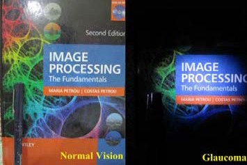
Figure 1. Normal vision vs. patient having glaucoma
Figure-1 shows how the objects are viewed by nor-
mal vision and patients having glaucoma.The different forms
of glaucoma are Open-angle glaucoma (OAG) ,Angle-closure glaucoma (ACG), Normal tension glaucoma (also called low tension glaucoma), Secondary glaucoma and Congenital glau- coma . The reasons may be family history of glaucoma , aged above 40 years, short-sighted, diabetic, a serious eye injury, using steroid treatment over an extended period or having hypertension.
In this paper preprocessing algorithms are compared
by the PSNR and MSE value. The paper is organized as fol- lows: Section II discusses the need for preprocessing. Section III presents the preprocessing techniques for eye fundus imag- es. The experimental evaluation is presented in section IV. Finally conclusion is given in section V.
II. NEED FOR PREPROCESSING
Retinal images are acquired with a digital fundus camera[1] which captures the illumination reflected from the retinal surface. Despite controlled conditions, many retinal images suffer from non –uniform illumination given by sever- al factrors: the curved surface of the retina, pupil dila- tion(highly variable among patients), or presence of diseases among others. The curved retinal surface and the geometrical configuration of the light source and camera lead to a poorly illuminated peripheral part of the retina with respect to the central part. Preprocessing can dramatically improve the per- formance of image processing methods like Image transform, Segmentation, Feature extraction and disease detection. Sev-
IJSER © 2013 http://www.ijser.org
International Journal of Scientific & Engineering Research, Volume 4, Issue 12, December-2013 1369
ISSN 2229-5518
eral techniques have been used to enhance retinal images.
III. PREPROCESSING METHODS FOR EYE FUNDUS IMAGES
Preprocessing is the step taken before the major im- age processing task. The problem here is to perform some basic tasks in order to render the resulting image more suita- ble for the job to follow. In this case it may involve enhancing the contrast, removing noise.
Preprocessing is the important step that influences automated detection of disease like glaucoma. The following are the preprocessing methods under study
(a) Contrast adjustment
(b) Adaptive Histogram equalization
(c) Average filtering
(d) Median filtering
(e) Homomorphic filtering
(a) Contrast adjustment
(b)Adaptive Histotram Equalization
Adaptive histogram equalization (AHE) is a computer image processing technique used to improve contrast in imag- es. It differs from ordinary histogram equalization in the re- spect that the adaptive method computes several histograms, each corresponding to a distinct section of the image, and uses them to redistribute the lightness values of the image. Figure-
3(a) shows the original image and 3(b) image after adaptive histogram equalization.
The contrast of an image is the distribution of its dark and light pixels. A low-contrast image exhibits small differ- ences between its light and dark pixel values. The histogram of a low-contrast image is narrow. Since the human eye is sensi-
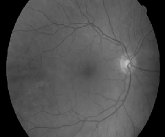
tive to contrast rather than absolute pixel intensities, a percep- tually better image could be obtained by stretching the histo- gram of an image so that the full dynamic range of the image is filled.Figure-2(a) shows the original and 2(b) the image after contrast adjustment.

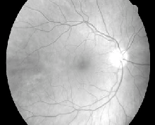
Figure 2(a). Original image
Figure 3(a). Original image
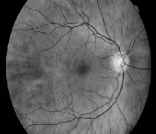
Figure 3(b). Adaptive histogram equalized image
(c) Average filtering
The Average (mean) filter[3] smooths image data, thus elimi- nating noise. This filter performs spatial filtering on each indi- vidual pixel in an image using the grey level values in a square or rectangular window surrounding each pixel.
For example:
a1 a2 a3
a4 a5 a6 3x3 filter window
a7 a8 a9
Figure 2(b). Contrast adjusted image
The average filter computes the sum of all pixels in the filter window and then divides the sum by the number of pixels in the filter window:
IJSER © 2013 http://www.ijser.org
International Journal of Scientific & Engineering Research, Volume 4, Issue 12, December-2013 1370
ISSN 2229-5518
Filtered pixel = (a1 + a2 + a3 + a4 ... + a9) / 9
Figure-4(a) shows the original image and 4(b) image after
Average filtering.


Figure 4(a). Original image
Figure 5(a). Original image

Figure 5(b). Median filtered image
(e) Homomorphic filtering
Homomorphic filter[6] is sometimes used for image enhancement. It simultaneously normalizes the brightness across an image and increases contrast. Here homomorphic filtering is used to remove multiplicative noise. Illumination and reflectance are not separable, but their approximate loca- tions in the frequency domain may be located. Since illumina-
tion and reflectance combine multiplicatively, the components

are made additive by taking the logarithm of the image inten- sity, so that these multiplicative components of the image can be separated linearly in the frequency domain. Illumination variations can be thought of as a multiplicative noise, and can be reduced by filtering in the log domain.
Figure 4(b). Average filtered image
(d) Median filtering
The Median Filter does somewhat the same, but instead of taking the mean or average, it takes the median. The median is gotten by sorting all the values from low to high, and then taking the value in the center. If there are two values in the center, the average of these two is taken. A median filter gives better results to remove salt and pepper noise, because it com- pletely eliminates the the noise.To get the median of the cur- rent pixel and it's 8 neighbors, set filterWidth and filterHeight to 3, but we can also make it higher to remove larger noise particles. Figure-5(a) shows the original image and 5(b) image after median filtering.
To make the illumination of an image more even, the

high-frequency components are increased and low-frequency components are decreased, because the high-frequency com- ponents are assumed to represent mostly the reflectance in the scene (the amount of light reflected off the object in the scene), whereas the low-frequency components are assumed to repre- sent mostly the illumination in the scene. That is, high-pass filtering is used to suppress low frequencies and amplify high frequencies, in the log-intensity domain[7]. Figure-6(a) shows the original image and 7(b) image after histogram equaliza- tion.

IJSER © 2013 http://www.ijser.org
International Journal of Scientific & Engineering Research, Volume 4, Issue 12, December-2013 1371
ISSN 2229-5518
Figure 6(a). Original image
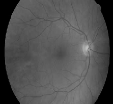
Figure 6(b). Homomorphic filtered image
IV. EXPERIMENTAL EVALUATION
To test the accuracy of preprocessing techniques , the fol- lowing three steps are used.
have a very wide dynamic range, PSNR is usually expressed in terms of the logarithmic decibel scale. A higher PSNR would normally indicate that the reconstruction is of higher quality
The PSNR is defined as:
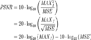
Here, MAXI is the maximum possible pixel value of the im- age.
• An eye fundus imagIe is takJen as input. SER
• Preprocessing technique is applied for fundus image.
• The MSE and PSNR value is calculated for different preprocessing techniques.
The PSNR and MSE values exhibit the performance of prepro- cessing techniques. To estimate the quality of the reconstruct- ed images, following parameters are used.
(a) Mean Square Error (MSE)
(b) Peak signal to Noise Ratio (PSNR)
(a)Mean Square Error (MSE)
Given a noise-free m×n monochrome image I and its noisy approximation K, the metric MSE is defined as:

Other metrics like Root Mean Square Deviation RMSE, Mean
Absolute Error MAE and PSNR are defined using MSE.
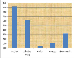
Figure 7.Comparison of different preprocessing algorithms by MSE
(b)Peak Signal to Noise Ratio (PSNR)
PSNR is the ratio between the maximum possible power of a signal and the power of corrupting noise that af- fects the fidelity of its representation. Because many signals
IJSER © 2013 http://www.ijser.org
International Journal of Scientific & Engineering Research, Volume 4, Issue 12, December-2013 1372
ISSN 2229-5518
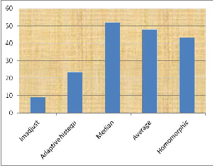
Figure8. Comparison of different preprocessing algorithms by PSNR
The figure-7 gives the MSE value for different image preprocessing algorithms and Figure-8 gives the PSNR ratio for different image preprocessing algorithms.From the fig- ures it can be concluded that median filtering technique gives high PSNR value and low mean square rate. So the median filtering technique gives desirable results compared to other preprocessing techniques.
V CONCLUSION
In the early detection of glaucoma the retinal image acquired using fundus camera is to be preprocessed before applying other image processing methods. The different pre- processing techniques like contrast adjustment, Adaptive his- togram equalization, Average filtering, Median filtering and Homomorphic filtering are applied to the fundus images in the gold standard database. These algorithms are evaluated using Peak Signal to Noise Ratio and Mean Square Error. The Median filtering and Average filtering give suitable results and Median filter is found to be better with high PSNR and low MSE values.
REFERENCES
IJSER
[1] D. Huang, P. K. Kaiser, C. Y. Lowder, and E. I.Traboulsi, Retinal
Imag ing. Mosby Elsevier, 2006.
[2] H. A. Quigely and A. T. Broman, “The number of people with glauo ma worldwide in 2010 and 2020,” BritishJournal of Oph thal mology, vol. 90, pp. 262–267, March2006. Hquig ley@jhmi.edu.
[3] Y. Wang, Q. Chen and B. Zhang. 1999. Image enhancement based on equal area dualistic sub-image histogram equalization method. IEEE Trans. on Consumer Electronics. 45(1): 68-75.
[4] http://m2matlabdb.ma.tum.de/files.jsp?MC_ID=12&SC_ID=18 [5]http://www.academia.edu/1569435/An_Introduction_to_Digital_Image_
processing_with_MATLAB [6]http://en.wikipedia.org/wiki/Homomorphic_filtering
[7] Douglas B. Williams and Vijay Madisetti (1999). Digital signal
processing Handbook. CRC Press. ISBN 0-8493- 2135-2.
[8] E. Sivalingam, “Glaucoma: An overview,” J Ophthalmic Nurs Tech
15(1), pp. 15–18, 1996.
[9] George R and Vijaya L (2008) ‘First world glaucoma day, march 6, 2008: Tackling glaucoma challenges in India’ Indian Journal of Ophthal mology, Vol.56, No.2, pp. 97-98.
[10] Anil K Jain, “Digital Image Processing”, 4th edition.
IJSER © 2013 http://www.ijser.org