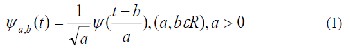
Where, a is the scale parameter and b the translation parame- ter. Practical implementation of wavelet transforms requires discretization of its translation and scale parameters by tak- ing,
Thus, the wavelet family can be defined as:
International Journal of Scientific & Engineering Research, Volume 4, Issue 7, July-2013 129
ISSN 2229-5518
An Overview of Different Image Fusion Methods for Medical Applications
Anjali A. Pure, Neelesh Gupta, Meha Shrivastava
Abstract— Image fusion is an important research topic in many related areas such as computer vision, remote sensing, robotics, and medical imaging etc. Image fusion is the process of combining relevant information from several images into single image. The final output image can provide more information than any of the single image. Now-a-days, almost all areas of medical diagnosis are impacted by the digital image processing. For medical diagnosis, MRI and CT images are of main concern, both images give special sophisticated characteristics of the organ to be imaged. Computed Tomography (CT) provides the best information on denser tissue like bones. Magnetic Resonance Image (MRI) provides better information on soft tissues. W ith more available multimodality medical images in clinical applications, the idea of combining images from different modalities become very important and medical image fusion has emerged as a new promising research field that help physicians in the diagnosis process. More research has been done for fusion of MRI and CT images using traditional wavelet transform and few attempts using curvelet transform. This paper provides an overview of different image fusion methods for medical applications in brief and to improve the fusion results proposed method is described shortly.
Index Terms— CT image, Curvelet Transform, Fast discrete curvelet transform (FDCT), Image fusion, MRI image, Wavelet Transfor
—————————— ——————————
Image fusion is a tool to combine multimodal images by us- ing image processing techniques. Specifically it aims at the integration of disparate and complementary data in order to enhance the information apparent in the images, as well as to increase the reliability of the interpretation [1],[2]. Due to the advent of new diseases complementary information are re- quired from different modalities. When sensitive organs like brain are scanned, both magnetic resonance imaging and computed tomography scans are preferred. CT provides best information about denser tissue and MRI offers better infor- mation on soft tissue [2],[3],[4]. These complementarities have led to idea that combining images acquired with different medical devices will generate an image that can offer more information than individual image. So, it is expected that fu- sion of MRI and CT images of the same organ would result in an integrated image of much more details [3]. Image fusion is deeply related to many different image processing fields such as satellite imaging, remote sensing and medical imaging. The study in the field of image fusion has evolved to serve the ad- vance in satellite imaging and then, it has been extended to the field of medical imaging. Several fusion algorithms have been proposed extending from the simple averaging to the curvelet transform [1],[3].
————————————————
1. Anjali A. Pure,ECE dept.,TIEIT, Bhopal, India. Email- pureanjali@gmail.com
2. Neelesh Gupta,HOD, ECE dept.,TIEIT, Bhopal, India.
3. Meha Shrivastava,Asst. Professor, ECE dept.,TIEIT, Bhopal, In dia.Email- mehakhare@gmail.com
Literature Survey reveals that, Shih-Gu Huang [1] proposed different image fusion methods that have been developed to perform image fusion. Wavelet transform used for image fu- sion produced superior results than different methods. S. Vasuki, S. Gandhimathi et al. [2] proposed integration of wavelets and PCA for fusion of medical images. Comparative analysis is done with different types of wavelets. The results obtained are suitable for medical image fusion. Smt. G. Ma- matha, L. Gayatri [3] proposed wavelet and curvelet transform for image fusion and comparison was done between these two transform. The results show superiority of curvelet transform than wavelet transform. A. Soma Sekhar, Dr. M. N. Giri Pra- sad [4] proposed integration of wavelet transform and PCA based image fusion for medical images and compare results with different types of wavelets, which gives better fusion results. Bharat and E.S Karthik Kumar [5] proposed imple- mentation of 2-G curvelet transform for image fusion which is fast and simpler than 1-G curvelet transform. Researchers have made lot of work on wavelet and curvelet transform for image denoising, image contrast enhancement, fusion of satel- lite images, image retrieval, texture analysis and object recog- nition. Few attempts were made for fusion of the MR and CT images using curvelet transform. This paper focuses more on image fusion for medical images. It was found that more work is done using wavelet transform for fusion of MRI and CT ap- plication. The traditional wavelets perform well only at repre- senting point singularities since they ignore the geometric properties of structures and do not exploit the regularity of edges.
The curvelet transform has evolved as a tool for the repre- sentation of curved shapes and regularity of edges. The wave- let transform applied to these two images preserve both spec- tral and spatial information and gives image details but it has limited directionality to deal with curved shapes. The solution to this is curvelet transform which has ability to deal with
IJSER © 2013 http://www.ijser.org
International Journal of Scientific & Engineering Research, Volume 4, Issue 7, July-2013 130
ISSN 2229-5518
curved shapes.
To fuse MRI and CT images there are many fusion algo- rithms based on Wavelet. Now in this paper, a new fusion method is proposed based on both wavelet and Second gener- ation Curvelet Transform. Since using Wavelet Transform edge information can’t be extracted clearly. In Medical Image Fusion the edge information is most important so a new method can be used to fuse these images. It’s a combination of Wavelet and Curvelet based algorithm which is used for fu- sion to extract edge information and the process is simple to analyze the fusion of the images. The performance of pro- posed method can be compare with different types of wavelets used in image fusion.
Wavelets are finite duration oscillatory functions with zero average value. The irregularity and good localization proper- ties make them better basis for analysis of signals with dis- continuities. Wavelets can be described by using two func- tions viz. the scaling function f (t), also known as father wavelet and the wavelet function or mother wavelet. Mother wavelet ψ (t) undergoes translation and scaling operations to give self-similar wavelet families as in (1).
Where, a is the scale parameter and b the translation parame- ter. Practical implementation of wavelet transforms requires discretization of its translation and scale parameters by tak- ing,![]()
Thus, the wavelet family can be defined as:![]()
.
The wavelets used in image fusion can be classified into three categories Orthogonal, Bi-orthogonal and A’trous wave- let. Although these wavelets share some common properties, each wavelet has a unique image decompression and recon- struction characteristics that lead to different fusion results [2],[ 6], [13].
the φj, k(x) and Ψj, k(x) are orthonormal, they include the fol- lowing property.
These results in a representation of a single image, containing multiscale detail information from all component images involved. This representation leads to multiple appli- cations ranging from multispectral image fusion to color and multivalued image enhancement, denoising and segmenta- tion.
2. Bi-orthogonal Wavelet
For biorthogonal transform, perfect reconstruction is available. Orthogonal wavelets give orthogonal matrices and unitary transforms; biorthogonal wavelets give invertible ma- trices and perfect reconstruction. For biorthogonal wavelet filter, the Low-pass and high-pass filters do not the same length. The low-pass filter is always Symmetrical, while high pass filter could be either symmetric or anti-symmetric. The method allows unusual flexibility in choosing a filter for any task involving the multiresolution analysis and synthesis.
3. A’trous (Non-orthogonal) Wavelet
A‘trous is a kind of non–orthogonal wavelet that is
different from orthogonal and biorthogonal. It is a stationary or redundant transform, i.e. decimation is not implemented during the process of wavelet transform, while the orthogonal or biorthogonal wavelet can be carried out using either deci- mation or undecimation mode. The enhancement of the spatial information often leads to the distortion of the information in the spectral domain.
The most common form of transform type image fusion algo- rithms is the wavelet fusion algorithm due to its simplicity and its ability to preserve the time and frequency details of the images to be fused. Wavelet transform fusion is more formally defined by considering the wavelet transforms of the two reg- istered input images together with the fusion rule .Then, the inverse wavelet transform is computed, and the fused image is reconstructed [1], [3], [5], [6] . 2-D DWT is very useful for im- age processing because the image data are discrete and the spatial and spectral resolution is dependent on the frequency. The DWT has the property that the spatial resolution is small in low-frequency bands but large in high frequency bands.
1. Orthogonal Wavelet
The dilations and translation of the scaling function
φj, k(x) constitute a basis for Vj, and Similarly Ψj, k(x) for Wj, if
IJSER © 2013 http://www.ijser.org
International Journal of Scientific & Engineering Research, Volume 4, Issue 7, July-2013 131
ISSN 2229-5518
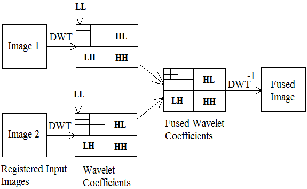
Fig.1 Block diagram of Discrete Wavelet transform
A schematic diagram of the wavelet fusion algorithm of two registered images I 1 (X1 , X2 ) and I 2 (X1 , X2 ) is depicted in fig.1. It can be represented by the following equation,
I(X1 , X2) =W-1{Ψ[W (I 1 (X1 , X2 )), W (I 2 (X1 , X2 ))]} (5)
Where W, W-1and ψ are the wavelet transform operator, the inverse wavelet transform operator and the fusion rule, re- spectively. There are several wavelet fusion rules that can be used for the selection of wavelet coefficients from the wavelet transforms of the images to be fused. The most frequently used rule is the maximum frequency rule which selects the coefficients that have the maximum absolute values. The wavelet transform concentrates on representing the image in multi-scale and it is appropriate to represent linear edges. For curved edges, the accuracy of edge localization in the wavelet transform is low. So, there is a need for an alternative ap- proach which has a high accuracy of curve localization such as the curvelet transform.
Candes and Donoho justifies that ,though wavelet trans- form exhibits time frequency localization and yields accepta- ble fused output, the edges and singularities are not well rep- resented. Also it suffers from limited directionality. The point singularity is better suited for wavelets in 1 dimensional sig- nals ,but 2 dimensional signals like images have curve or line singularities where wavelets fails to approximate. Hence to process images of sparse nature, Fast Discrete curvelet trans- form is recommended by the researchers. E. J. Candes and D. L. Donoho put forward curvelet transform theory in 2005[7]. The curvelet transform is a multiscale directional transform that allows an almost optimal nonadaptive sparse representa- tion of objects with edges. Curvelet transform consisted of special filtering process and multiscale Ridgelet transform. Curvelet transform is anisotropy, which can represent the con- tour of image more sparsely and provide more information for image processing. The curvelet transform has gone through two major revisions. The first generation curvelet transform used a complex series of steps involving the ridgelet analysis
of radon transform of an image. In this curvelet approach, in- put image is first decomposed into a set of sub bands each of which is then partitioned into several blocks for ridgelet anal-
ysis. The ridgelet transform is implemented using the Radon transform and the 1-D wavelet transform. During the ridgelet transform, one of the processes is the spatial partitioning which involves overlapping of windows to avoid blocking effects. It results in a large amount of redundancy. The Curvelet Co-efficient has taken and depending upon the max- imum value the fusion between the images takes place. It is used to extract the edge information, the ridgelet transform is complicated so the process is very time consuming and the performance was exceeding slow.
The second generation curvelet transform discarded the use of the ridgelet transform, thus reduced the amount of redundan- cy in the transform and increased the speed considerably. Two fast discrete curvelet transform (FDCT) algorithm were intro- duced. The first algorithm is based on unequally-spaced FFT while the second is based on the wrapping of specially select- ed Fourier samples. In this paper, we focus on the “wrapping” version of the curvelet transform.
The algorithm can be summarized as follows-![]()
1. Apply the 2D FFT and obtain Fourier samples![]()
2. For each scale j and angle l form the product![]()
3. Wrap this product around the origin and obtain
Where, the range for n1 and n2 is now 0 < n1 < L1,j and 0 < n2
< L2,j (for θ in the range (−π/4, π/4)).
4. Apply the inverse 2D FFT to each ˜ fj,l, hence collecting the
discrete coefficients cD(j, l, k).
It is clear that this algorithm has computational complexity
O (n2 log n).
The wavelet transform concentrates on representing the image in multi-scale and it is appropriate to represent linear edges. For curved edges, the accuracy of edge localization in the wavelet transform is low. Normally, when a wavelet trans- formation alone is applied the results are not so useful for analysis. For medical applications, MRI and CT images plays very important role. Since medical images have several objects and curved shapes. So, there is a need for an alternative ap- proach which has a high accuracy of curve localization, it is expected that the curvelet transform would be better in their fusion.
Wavelet transform is useful for objects with point singu- larities and analyses the feature of images in detailed, but it
IJSER © 2013 http://www.ijser.org
International Journal of Scientific & Engineering Research, Volume 4, Issue 7, July-2013 132
ISSN 2229-5518
does not provide information about edges clearly. While curvelet transform is more useful for the analysis of images having curved shape edges. So, in this paper, a new image fusion technique based on the combination of wavelet and a fast discrete curvelet transform is proposed, which describe the curved shapes of images and analyses feature of images better. In medical image fusion, edges play a very important role. This new fusion technique is used to fuse the MRI and CT images. These two images contain complementary infor- mation. The Flow of proposed image fusion method can be given as follows: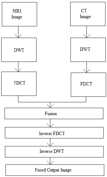
Fig. 2 Flow of proposed image fusion method
In many important imaging applications, images exhibit edges and discontinuities across curves. In biological imagery, this occurs whenever two organs or tissue structures meet. Espe- cially in image fusion the edge preservation is important in obtaining the complementary details of the input images. As edge representation in curvelet transform is better and wave- let transform gives image details. The combination of wavelet and fast discrete curvelet transform applied to medical images may give better fusion results useful for diagnosis. This can be used to give the edge information clearly and speed of compu- tation will be high compare to other methods. The proposed method may be useful for researchers for further research work on image fusion.
[1] Shih-Gu Huang, Wavelet for Image Fusion.
[2] S.Vasuki, S. Gandhimathi, S. Manika VInodhini,’ Comparative
Analysis of Wavelets for Fusion Application’, IJCA, 2012.
[3] Smt.G.Mamatha,L.Gayatri,’AN IMAGE FUSION USING WAVE- LET AND CURVELET TRANSFORMS’, Global Journal of Ad- vanced Engineering Technologies,Vol1,Issue-2,2012,ISSN:2277-
6370.
[4] A. Soma Sekhar, Dr.M.N.GiriPrasad, ’A Novel Approach of Image
Fusion on MR and CT Images Using Wavelet Transforms, IEEE
2011.
[5] S.Bharath and E.S. Karthik Kumar, Implementation Of Image Fu- sion Algorithm Using 2gcurvelet Transforms, ISBN 978-1-4675-
2248-9@2012.
[6] Bin Yang and Shutao Li, Multifocus Image Fusion and Restoration with Sparse Representation, IEEE 2010.
[7] Emmanuel Cand`es, Laurent Demanet, David Donoho and Lexing Ying; Fast Discrete Curvelet Transforms,Applied and Computa- tional Mathematics, Caltech Pasadena 2006.
[8] Jianwei Ma and GerlindPlonka, The Curvelet Transform, IEEE SIGNAL PROCESSING MAGAZINE[118] MARCH 2010.
[9] Gang Hong, Yun Zhang,’The effect of different types of wavelets on image fusion.
[10] Deepa M, Wavelet and Curvelet Based Threshholding Techniques for Image Denoising, IJARCSEE vol1 Issue2012,ISSN:227-9043.
[11] E.J.Candes, D.L.Donoho Curvelets: A Surprisingly Effective Non- adaptive Reprentation for Objects with Edges.
[12] M. Sifuzzaman M.R. Islam,M.Z.Ali, Application Of Wavelet and
its Advantages Compared to Fourier Transform,Journal of Physi- cal Science,Vol.13,2009,121-134,ISSN:0972-8791.
[13] Y.Kiran Kumar, Comparison of Fusion Techniques Applied to pre- clinical images: Fast Discrete Curvelet Transform using Wrapping Technique & Wavelet Transform, JATIT2009.
[14] Pao-Yen Lin, An introduction wavelet transforms.
IJSER © 2013 http://www.ijser.org
International Journal of Scientific & Engineering Research, Vo lume 4, Issue 7, July-2013
ISSN 2229-5518
133
[15] Myungjin Choi, Rae Young Kim, Moon-GyuKim,'The curvelet transform for image fusion.
[16] Rafael C. Gonzalez and Richard E. Woods, Digital image Pro
cessing, Third Edition.
IJSER lb)2013