International Journal of Scientific & Engineering Research, Volume 5, Issue 9, September-2014 67
ISSN 2229-5518
Agenesis of maxillary primary and permanent lateral incisor
Rena Aphraim, Shubha M.
Abstract— Dental agenesis is one of the most common developmental anomaly in humans and is many a times associated with several other oral abnormalities. Hypodontia is uncommon in the deciduous dentition with a prevalence that ranges from 0.5% to 0.9%, with the maxillary lateral incisor being the most affected unilaterally or bilaterally. The absence of a deciduous tooth is associated strongly with an increased prevalence of a missing succedaeous tooth. A tooth is defined to be congenitally missing if it has not erupted into the oral cavity and has not been extracted or accidentally lost or is found missing in the radiograph. A disturbance during the early stages of tooth development can result in its congenital absence. Hypodontia can occur either as an isolated condition involving one tooth, a few or many teeth or can be associated with a condition or syndrome essentially reflecting the genetically and phenotypical heterogenecity of the condition. It is also more frequently observed in females than in males. In primary dentition early diagnosis of missing anterior teeth is not usual, and might go undiagnosed till the age of 12 years, till aesthetic treatment is sought, for missing permanent maxillary incisors. Hobkirk et al reported that in 451 patients treated for hypodontia, more than 50% were older than 12 years and all reported f or aesthetic or prosthetic rehabilitation.An early x-ray examination, using periapical and panoramic techniques, and if necessary computed tomography, may be necessary to correctly diagnose the particular situation and get information regarding the treatment modalities. The delay in diagnosis and treatment of this condition can lead to an unpleasant smile, facial asymmetry, midline diastema,dental arch discrepancy,occlusal disharmony, canine impactions, decreased periodontal health and pschycoligical conditions. The objective of this article is to bring awareness among the dentists and to stress upon the importance of early diagnosis of missing lateral incisors in the early mixed dentition period, its consequences and the possible earliest management during this period to restore aesthetics and function to the affected patient.
Index Terms— Dental Agenesis, Hypodontia, Lateral incisor, Pedodontics.
—————————— ——————————
1 INTRODUCTION
ental agenesis is one of the most common developmental anamoly in humans and is many a times associated with several other oral abnormalities. Hypodontia is uncom-
mon in the deciduous dentition with a prevelance that ranges from 0.5% to 0.9%, with the maxillary lateral incisor being the most affected unilaterally or bilaterally. The absence of a de- ciduous tooth is associated strongly with an increased preva- lence of a missing succedaeous tooth. A tooth is defined to be congenitally missing if it has not erupted into the oral cavity and has not been extracted or accidentally lost or is found missing in the radiograph. A disturbance during the early stages of tooth development can result in its congenital ab- sence. Hypodontia can occur either as an isolated condition involving one tooth, a few or many teeth or can be associated with a condition or syndrome essentially reflecting the genet- ically observed in females than in males[1]
In primary dentition early diagnosis of missing anterior teeth is not usual, and might go undiagnosed till the age of 12 years, till aesthetic treatment is sought, for missing permanent maxillary incisors. Many studies have confirmed the associa- tion of agenesis of maxillary lateral incisors with other tooth anomalies. Patients with maxillary lateral agenesis has a sig-
————————————————
• Rena Aphraim is currently Professor, Department of Pedodontics and Preventive Dentistry, Mahe Institute of Dental Sciences, Mahe, UT of Pu- dussery, India
• Shubha M is currently Reader, Department of Pedodontics and Preventive
Dentistry, Mahe Institute of Dental Sciences, Mahe, UT of Pudussery, In-
dia.
nificantly increased prevalence rate of 18.2% in permanent tooth agenesis.[2] Ectopic eruption of canines, or canine im- pactions were considerably increased in incidence with agene- sis of maxillary lateral incisor. Generalized microdontia was seen as a variable expression of agenesis in patients. De- creased periodontal health associated with maxillary lateral incisor agenesis is also of concern to the pediatric dentist.[3]
Hobkirk et al reported that in 451 patients treated for hy- podontia, more than 50% were older than 12 years and all re- ported for aesthetic or prosthetic rehabilitation.[4] The objec- tive of this article is to bring awareness among the dentists and to stress upon the importance of early diagnosis of miss- ing lateral incisors, its consequences and the possible earliest management to restore aesthetics and function and to render the best possible treatment to the affected patient..
2 CASE REPORT
A patient aged8 years reported to the department of Pedo- dontics with the complaint of aesthetically displeasing exces- sive spacing in the midline and unerupted permanent maxil- lary lateral incisors .On clinical examination the erupted max- illary central incisors showed considerable median diastema, of 4-5mm and the lateral incisors were missing in the oral cavi- ty.(Fig-1) A detailed history was taken to rule out the possibil- ity of extraction or accidental loss of the primary lateral inci- sors , or the presence of any associated syndromes . The oral status of the patient was satisfactory and the gingival tissues appeared to be clinically of optimal health.The patient howev- er did not report for further treatment due to personal reasons. She reported to us at the age of 11 years for the treatment of
IJSER © 2014 http://www.ijser.org
International Journal of Scientific & Engineering Research, Volume 5, Issue 9, September-2014 68
ISSN 2229-5518
her median diastema. The canines had been extracted from elsewhere to accommodate her lateral incisors, which failed to erupt to date. She was referred to us for a thorough investiga- tion and further treatment. Orthopantomogram showed the central incisors exhibiting considerable diastema of the crowns and roots andthe bilateral absence of maxillary lateral inci- sors.(Fig-2) The other teeth in the dentition seemed to be healthy and erupting satisfactorily. The maxillary canine bilat- erally was present and showed no signs of impaction or altera- tion in the path of eruption. There was however agenesis of the third molars. Dental age of the patient was considerably delayed. A provisional diagnosis of congenital absence of bi- lateral maxillary lateral incisors was made which resulted in considerable midline diastema. A multidisciplinary approach of management was planned, due to the complexity of the situation and necessity ofa long duration of treatment. Treat- ment consisted of orthodontic, surgical, andaprosthodontic intervention. The treatment protocol was explained to the par- ents and their informed consent obtained .Orthodontic treat-
permanent antagonists, and microdontia can occur in the suc- cedaneous permanent dentition which can result in an unaes- thetic appearance.[5] Radiographs must be taken at the earliest to confirm diagnosis of missing succedaneous teeth. Or- thopantomogram is the x-ray of choice as suggested by Pilo et al for an early diagnostic procedure in patients younger than 8 years.[6] In the event of agenesis of a primary lateral incisor and its successor, the paradoxical frequency of a malpostioned maxillary permanent canine shows the importance these teeth have as the guiding tooth in the eruption of the canines. [7,8] Maxillary canines have been believed to take the support of the root of the lateral incisor for guidance to erupt into the primary position. Hence its absence or malformation may re- sult in deviation in its path. Genetic factor also has been at- tributed to the deviation of the path of its eruption.[3,9]
Patients with agenesis of maxillary lateral insicors had a sig- nificantlyincreased prevalence rate of permanent tooth agene- sis of 18.2%, agenesis of 3rd
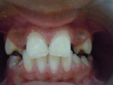
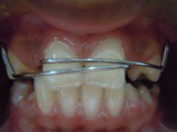
ment commenced by the closure of the midline diastema with
the help of a removable appliance incorporated with a split
labial bow.(Fig-3 & Fig-4) Considering the young age of the
patient closure of diastema was followed by the placement of
a temperory removable functional prosthesis to restore aes-
thetics.(Fig-5) A periodic follow up of the patient would be
necessary for a long period of time. The maxillary canines
would have to be guided to the position of the missing lateral
incisor and its recontouring done, with composite or crown, to
resemble a lateral incisor, after its eruption. The second option
would be the placement of an implant prosthesis on the eden-
tulous ridge spaces after proper evaluation, at a later date.

Figure 5.
molars, class II malocllusion,
and overretention of mandib-
ular central incisor.[4,10]
However our patient exhibit-
ed no other anomaly than
agenesis of the third molars.
The assessment of the golden
proportion in the facial view,
that is, the tooth-tooth width
proportion was found to be
missing in majority of the
cases treated with agenesis of
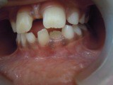
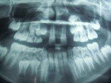
Figure 1. Figure 2.
Figure 3. Figure 4.
3 DISCUSSION
Detailed examination is essential for the diagnosis of missing lateral incisors in the early primary dentition. With the erup- tion of the permanent teeth in the early mixed dentition asymmetric loss of primary teeth,midline diastema, tooth mi- gration, midline shift to the affected side, over eruption of the
maxillary lateral incisor.[11].This was however not true in the situation encountered by us. The mesiodistalwidth of the re- maining anterior teeth were found to be almost of normal val- ues. The need for early recognition of the condition and its early intervention becomes extremely significant in the wake of these clinical signs.
4 CONCLUSION
There has been reported to be a close association between agenesis of the permanent maxillary lateral incisor and other tooth anamolies such as microdontia of the permanent maxil- lary lateral incisor, both in the individual and other relatives in a study.[3,10,12] Third molar absence is significantly more frequent in individuals with agenesis of the permanent maxil- lary lateral incisor as was evident in our patient.Influenced by several factors interacting at different levels a common genetic mechanism might be evidently controlling this phenomena. [3,13] Early diagnosis and effective clinical management of missing lateral incisor is hence very important as itcan affect aesthetics and function and can pschycologically affect the child’s personality.
REFERENCES
[1] Textbook of Oral and Maxillofacial pathology by S. Neville, Damn. Allen, Bouquot.pg 77-80. 3rdedition.Elsevier Publications, a division of Reed Elsevier India Pvt ltd.
IJSER © 2014 http://www.ijser.org
International Journal of Scientific & Engineering Research, Volume 5, Issue 9, September-2014 69
ISSN 2229-5518
[2] PJ De Coster, LA Marks, LC Martius, AHuysenne. Dental agenesis: genetic and clinical perspectives. J Oral Path Med Jan 2009;38:1:1-17.
[3] Pinho T, Tavares P,MacielP, Pollmann C. Developmental disturbance of maxillary anterior lateral incisor in Portugese population. Eur J Orthod
2005;27:443-449.
[4] Hobkirk JA, Goodman JR, Jones SP. Presenting complaints and findings in a group of patients attending a hypodontia clinic. Br Dent J 1994;177:337-339.
[5] Bergendal B, Bergendal T, HallostenA, Koch G et al.A multidisciplinary ap-
proach to oral rehabilitation with osseointegrated implants in child and ado- lescents with multiple aplasia.Eur J Orthod:1996;18:119-129.
[6] Pilo, R, Kaffe,I et al. Diagnosis of developmental anomalies using panoramic radiograph. ASDC Journal Dentistry for children 54(4):267-272.
[7] Peck S, Peck L, Kataja M. Concomittant occurrence of canine malposition and tooth agenesis: evidence of genetic orofacial fields. Am J OrthodDentofacial Or-thop2002;122:657-660
[8] Al Nimri, Kazem S, Broul, Enas. Maxillary palatal canine impaction develop- ing in subjects with congenitally missing Maxillary Lateral Incisor. Am J Or- thodDemtofacialOrthop.Jul 2011;140(1):81-86.
[9] Hobkirk JA, Goodman JR, Jones SP. Presenting complaints and findings in a group of patients attending a hypodontia clinic. Br Dent J 1994;177:337-339.
[10] Garib DG, Alencar BM, Lauris JR, Bacetti T. Agenesis of Maxillary Lateral Incisor and associated dental anomalies. Am J OrthodDentofacialOrthop 2010 jun;237(6):732-735.
[11] Pinc, NublaPaveri, De Marchi et al. Analysis of the golden proportion and width/height ratios of maxillary anterior dentition in patients with lateral in- cisor agenesis. J Esthetic and Restorative Dentistry:2012.vol14(6)402-414.
[12] Yaquob, OmarDiBiase, Andrew Tetal. Mesiodistal tooth size relationship between bilateral congenital absence of Maxillary Lateral Incisor and anterior tooth width. Am JOrthoddentofacial Orthop.Mar2011;139(3):229-233.
[13] PJ De Coster, LA Marks, LC Martius, AHuysenne. Dental agenesis: genetic
and clinical perspectives. J Oral Path Med Jan 2009;38:1:1-17.
IJSER © 2014 http://www.ijser.org