International Journal of Scientific & Engineering Research, Volume 3, Issue 6, June-2012 1
ISSN 2229-5518
A Proposed technique for Brain MRI Using Region
Based Segmentation
Harman Kataria, Alka Jindal
————————————————————
Medical imaging plays an important role in patient diagnosis. To examine the disease of patient imaging technology is used. MRI and PET/CT is vital diagnostic imaging techniques. MRI and PET/CT provides a good contrast between the different soft tissues of the brain. Combined PET/CT instrumentation is a new type of advanced medical imaging equipments which is a high- performance combination of PET and CT scanners [1].
Segmentation is a challenging task. Segmentation helps in diagnosis of various diseases. We can apply research on the segmented data such as classifying a voxels, volumetric analysis of particular tissues and structures.
We can categorize the active contour model into two parts edge based models [2] and region based models [3]. This paper presents the segmentation methods Improved Level Set[4], Local Region based active contour[5] and variational level set method[6] and proposed model for extracting the abnormal tissue from the brain MRI images.
This paper presents the method that applies on both homogenous and non homogenous method. Intensity homogeneity occurs due to technical limitations or an artifact introduced by the object and it is mainly present in real images. The radio frequency coils produces nonuniform magnetic field that arise inhomogenity in magnetic resonance image (MRI). We require segmentation method to correct the intensity inhomogenity as a pre- processing step [7].
The paper is organized as follows. In section II we present different image segmentation methods. Section III describes
the experimental results and discussion Section IV describe the comparison of all segmentation method Section V
conclude the paper
Segmentation of brain is very important for doctors to make a definite diagnosis. The segmentation methods for Brain MRI are:
The author Jia Di et al[4], proposed a C-V model that makes segmentation better than the other model. Active contour model is used to evolve the curve on the given image in order to detect objects in the image. C-V model divides the entire region into two parts one is inside part and the other is outside part and we can get the edges from the end of curves iteration. The energy function is following as![]()
![]()
![]()
![]()
![]()
22![]()
(1)
There are two energy functions internal and external energy and each iteration should compute internal average gray c1 and external gray c2, and for solution we use Heaviside function![]()
![]()
![]()
![]()
![]()
12 (2)
IJSER © 2012 http://www.ijser.org
International Journal of Scientific & Engineering Research, Volume 3, Issue 6, June-2012 2
ISSN 2229-5518
![]()
![]()
![]()
![]()
(4)
Local Region Based Active Contour is a region based method that is insensitive to noise and it is robust against the initial curve placement. It segments non-homogenous objects. It uses smaller local regions to segment the object rather than the global region [5]. Three types of energies can be used with this methods uniform modelling energy, mean separation energy and histogram separation energy [8]. The final energy is shown as below:![]()
![]()
+ ![]() (5)
(5)![]()
![]()
![]()
A(x,y)= (6)![]()
(7)![]()
![]()
(8)
Where ![]() are the mean intensities of the interior and exterior regions.
are the mean intensities of the interior and exterior regions. ![]() is dirac function,
is dirac function, ![]() Force function,
Force function, ![]() is input image.
is input image.
The author Chunming Li et.al[6] proposed a Variational level set formulation method that is a region based method. This algorithm segments the whole image. It overcomes the problem that occurred due to intensity inhomogeneity. In this method we first define Region Scalable fitting (RSF)
energy function and localization is performed by two fitting
function f1, f2 and ![]() . This algorithm is able to segment
. This algorithm is able to segment
inhomogeneous objects. The standard gradient descent method is use to minimize the energy functional. The minimization equation is.![]()
![]()
![]()
E( ) ![]() –
– ![]()
![]()
![]()
![]()
![]()
![]()
(9)
Where I(x) is the image intensity at pixel x, H is a Heaviside function, ![]() is a Gaussian kernel defined as:
is a Gaussian kernel defined as:![]()
(10)![]()
With a scale parameter >0, f1 and f2 are two functions centred at pixel as![]()
![]()
(11)![]()
![]()
(12)
![]() are two constants that have been set 5 in the interface. The evolution equation is defined as below.
are two constants that have been set 5 in the interface. The evolution equation is defined as below.
![]()
![]() +
+
![]()
![]()
![]()
![]()
) − 1 2![]() )+ ( + (∇2 ( − )) (13)
)+ ( + (∇2 ( − )) (13)
![]() is a signed distance function.
is a signed distance function.
The first two terms is a data fitting term [9], these terms are used for driving active contour toward object boundaries. The third and fourth terms are used for smoothening and maintain the regularity of the contour.
————————————————
![]() Harman Kataria is currently pursuing masters degree program in Computer science engineering in PEC University of Technology, INDIA, E-mail: hrmn.kataria@gmail.com
Harman Kataria is currently pursuing masters degree program in Computer science engineering in PEC University of Technology, INDIA, E-mail: hrmn.kataria@gmail.com
IJSER © 2012 http://www.ijser.org
International Journal of Scientific & Engineering Research, Volume 3, Issue 6, June-2012 3
ISSN 2229-5518
The proposed model, is a variation of variational set level,
peculiarly. We can extract the tumour itself without the background of the image. This can be done with inclusion of properties like solidity and eccentricity. The proposed model can also be applied on nonhomogenous images. The model takes into account in reduction of iterations and hence increases in the speed.
In order to test the performance and discuss the comparison between the Improved Level Set Method [4], Local Region Based Active Contour [5] and Variational Level Set Formulation [6] and the proposed model. In this paper these algorithm has been tested with synthetic images of T2
Weighted brain MRI. The discussed algorithms were
implemented in MATLAB. We use the various parameters for comparisons viz., the no of iterations, intensity homogeneity and time taken by the algorithm to segment the brain MRI image. All these methods applied on a noisy synthetic image. The phenomenon of pre-processing and post processing is avoided so that we can compare the results precisely.
All these methods accurately segment the tissues and all methods are independent of the location of the initial contour. The first method is based on images with homogenous foreground and background. The second and third method is based on both non-homogenous foreground and background images.
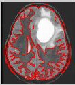
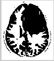
A Comparison of these algorithms is discussed in the above table. The improved level set method [4] should be applied to homogenous foreground and background images in Fig.2(c) it does not segment the tumour portion. Local Region Based active contour [5] accurately segment the image shown in Fig.3(c) but the problem is to initialize the contour for each and every image. Variational Level Set formulation [6] segment the whole image and this method can accurately show the tumour part in Fig.4(c). Number of iteration of these algorithms is given in table 1. The
proposed model used the properties like scalability and
eccentricity to reduce the no of iterations and increase the
speed of the model. The proposed model applied on both homogenous and nonhomogenous brain MRI images.
Table 1 Comparison of Segmentation Techniques
g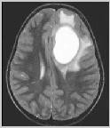
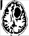
Fig. 1 Original T2 Weighted Brain MRI image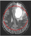
(a) (b) (c)
IJSER © 2012 http://www.ijser.org
International Journal of Scientific & Engineering Research, Volume 3, Issue 6, June-2012 4
ISSN 2229-5518
Fig.2 Results of the Improved Level Set Method (a)
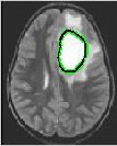
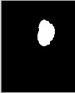
Initialization (b) Final contour 200 iterations (c) Final segmentation.
(a) (b) (c)
Fig.3 Results of the Local Region Based Method (a) Initialization (b)Final Contour 430 iterations (c) Final segmentation.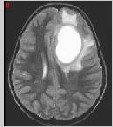
(a) (b) (c)
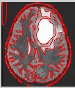
Fig.3 Results of the Variational Level Set Formulation (a) Initialization (b) Final Contour 150 iterations (c) Final segmentation.
In this paper, various region based segmentation algorithm for brain MRI such as Improved Level Set, Local Region based active contour and Variational Level Set formulation has been discussed. Amongst all the segmentation method discussed Variatonal Level Set Formulation is the best segmentation method in terms of the classification of the tissue recognition and tumour extraction. It clearly segments the whole image and can be applied to both homogenous and non homogenous images. In this method there is no need to initialize the contour. In localized Level
Set method the problem is to initialize the contour.
Improved Level Set method should be applied to only
homogenous images.
[1] Wang Rongfu, PET/CT Tumour Diagonsis, 1st ed., Peking University Medical Press, Beijing, 2007, pp.3-13.(in Chinese)
[2] V.Caselles, R. Kimmel, and G. Sapiro,”Geodesic active contour”,Int. J. Comput. Vis, vol. 22, pp.61-79, 1997.
[3] T. Chan and L. Vese,” Active contour without edges,”
IEEE trans Image Process., vol. 10, no. 2, pp 266-277, Feb.
2001.
[4] Jia Di, Yang Jin-Zhu, Zhang Yi-Fei. “An Efficient Modified Level Set Method For Brain Tissue Segmentation”, IEEE International Conference on Information and Automation june 2010.
[5]. Shawn Lankton, Allen Tannenbaum, “Localizing Region-Based Active Contours,” IEEE TRANSACTIONS ON IMAGE PROCESSING, VOL. 17, NO. 11, NOVEMBER
2008.
[6] Chunming Li ,Chiu-Yen Kao and John C.Gore,”Minimization of Region-Scalable Fitting Energy for Image Segmentation”,IEEE Trans. On Image Processing, vol.17,n0. 10,October 2008.
[7] Z. Hou,”A review on mr image intensity inhomogenity correction,”Int. J. Biomed. Imag.,2006.
[8]. J. A. Yezzi, A. Tsai, and A.Willsky, “A fully global approach to image segmentation via coupled curve evolution equations,” J. Vis. Comm. Image Rep., vol. 13, no. 1, pp. 195–216, Mar. 2002.
[9] L. Vese and T. Chan, “A multiphase level set framework for image segmentation using the Mumford and Shah model, ”Int.J.Comput.Vis.,vol. 50, pp. 271–293, 2002.
IJSER © 2012 http://www.ijser.org